info@c-tech-implant.com +39 051 66 61 817 + 49 721 60 95 32 38

    • Italiano
    • English
    • Deutsch
    • Español
    • Français
    • Русский
    • 中文 (中国)
    • Türkçe
    • Ελληνικά
    • Magyar
    • Српски језик
    • Lietuviškai
    • Bahasa Indonesia
    • Slovenčina

Slide Pubblicazioni Scientifiche

Scientific Publications

International Journal of Implant Dentistry – August 2017 – Investigation of peri-implant tissue conditions and peri-implant tissue stability in implants placed with simultaneous augmentation procedure: a 3-year retrospective follow-up analysis of a newly developed bone level implant system

Written by feRKzzCTo0 on . Posted in Bone Regeneration, Century-Gtr, Conventional Implantology, El – Esthetic Line, Henriette Lerner, Robert Sader, Scientific Publications, Shahram Ghanaati

International Journal of Implant Dentistry • August 2017

Investigation of peri-implant tissue conditions and peri-implant tissue stability in implants placed with simultaneous augmentation procedure: a 3-year retrospective follow-up analysis of a newly developed bone level implant system

Jonas Lorenz University Hospital Frankfurt · Department of oral, maxillofacial and plastic surgery, Henriette Lerner HL DENTCLINIC, Robert A. Sader Goethe-Universität Frankfurt am Main · Center of Stomatology
and Shahram Ghanaati Goethe University of Frankfurt/Main; Universitätsmedizin der Johannes Gutenberg-Universität Mainz · Department for Oral, Craniomaxillofacial and Facial Plastic Surgery; Institute of Pathology

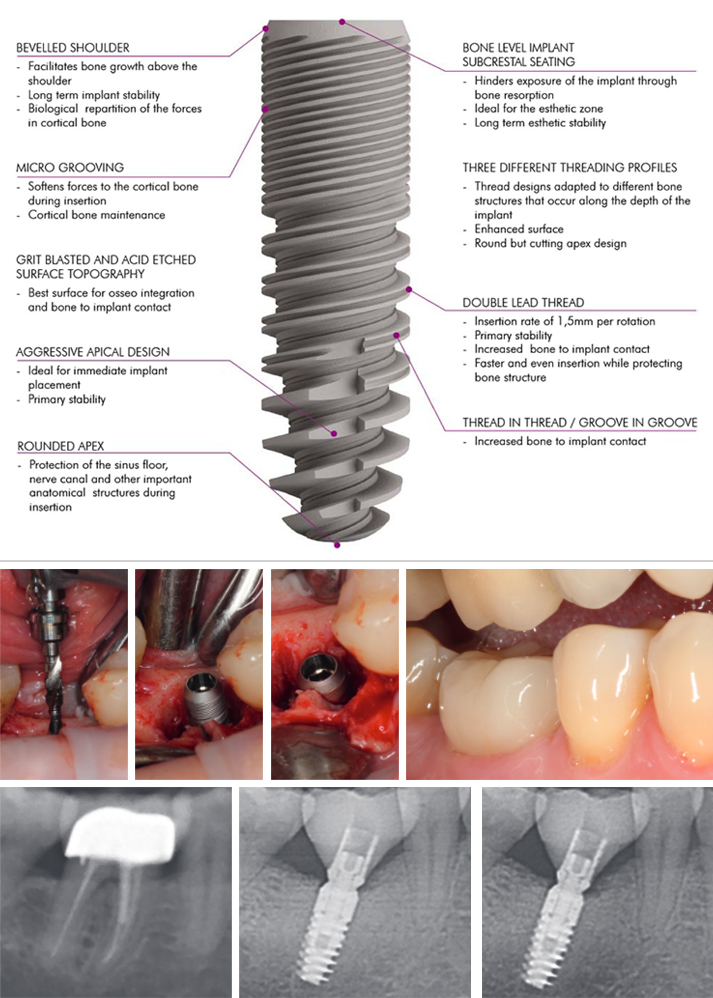
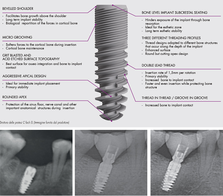
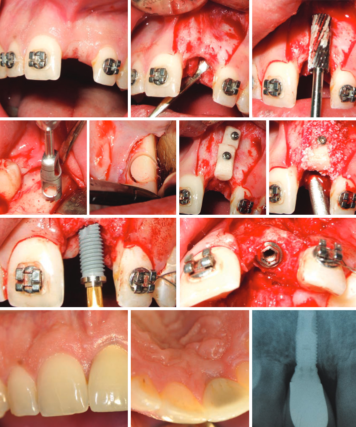
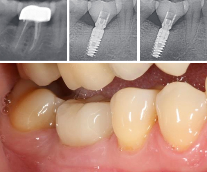
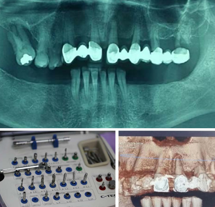
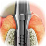
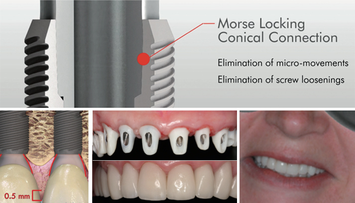
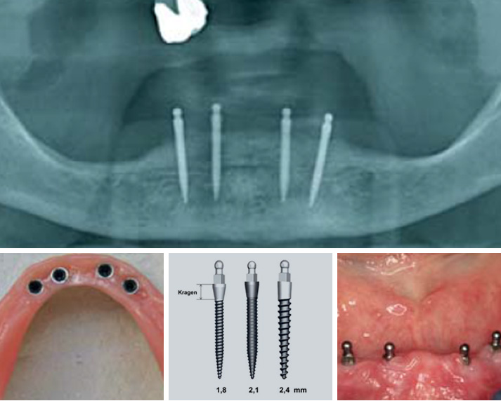
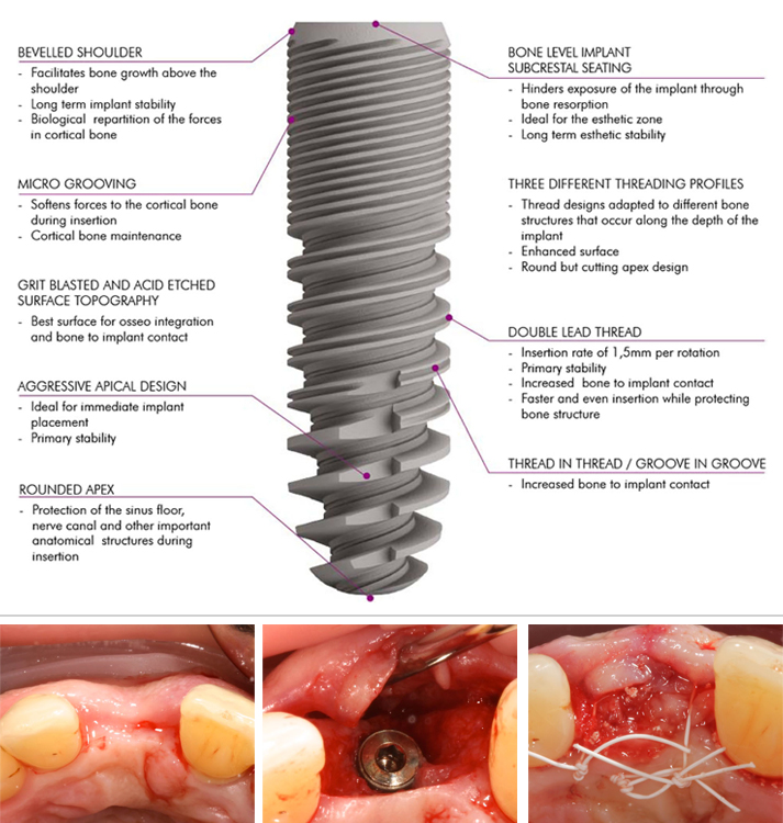
The aim of the present retrospective analysis was to assess peri-implant tissue conditions and document peri-implant tissue stability in C-Tech implants when placed simultaneously with a GBR (Guided Bone Regeneration) augmentation procedure.

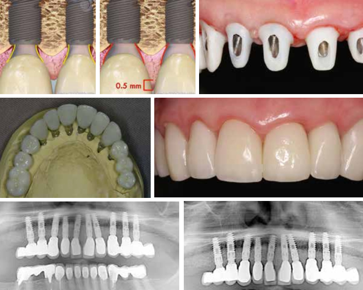
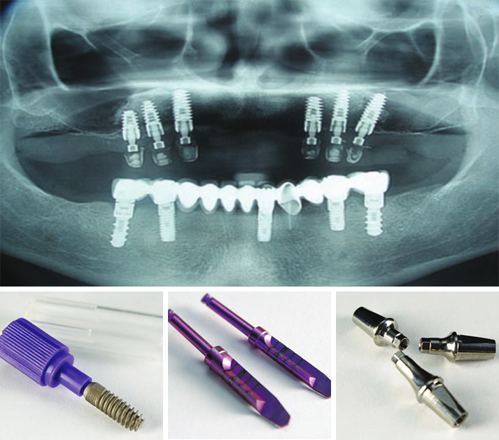
A total of 47 implants, which were placed simultaneously with a GBR (Guided Bone Regeneration) procedure with a synthetic bone substitute material in 20 patients, were investigated clinically and radiologically at least 3 years after loading.

The follow-up investigation revealed a survival rate of 100% and only low median rates for probing depths (2.7 mm) and BOP (bleeding on probing) (30%). The mean PES (Pink Esthetic Score) was 10.1 from the maximum value of 14.
No osseous peri-implant defects were obvious, and the mean bone loss was 0.55 mm.

In conclusion, implants placed in combination with a GBR (Guided Bone Regeneration) procedure can achieve long-term stable functionally and esthetically satisfying results for replacing missing teeth in cases of atrophy of the alveolar crest.


References

1. Gurgel BC, Montenegro SC, Dantas PM, Pascoal AL, Lima KC, Calderon PD. Frequency of peri-implant diseases and associated factors. Clin Oral Implants Res. 2016; doi: 10.1111/clr.12944

2. Qian J, Wennerberg A, Albrektsson T. Reasons for marginal bone loss around oral implants. Clin Implant Dent Relat Res. 2012;14(6):792–807.

3. Berglundh T, Lindhe J, Ericsson I, Marinello C, Liljenberg B, Thomsen P. The soft tissue barrier at implants and teeth. Clin Oral Implants Res. 1991;2:81–90.

4. Berglundh T, Lindhe J, Jonsson K, Ericsson I. The topography of the vascular systems in the per iodontal and peri-implant tissues in the dog. J Clin Periodontol. 1999;21:189–93.

5. Moon I, Berglundh T, Abrahamsson I, Linder E, Lindhe J. The barrier between the keratinized mucosa and the dental implant. An experimental study in the dog. J Clin Periodontol. 1999;26:658–63.

6. Lindhe J, Berglundh T. The interface between the mucosa and the implant. Periodontol. 1998;17:47–54.

7. Masaki C, Nakamoto T, Mukaibo T, Kondo Y, Hosokawa R. Strategies for alveolar ridge reconstruction and preservation for implant therapy. J Prosthodont Res. 2015;59(4):220–8.

8. Damien CJ, Parsons JR. Bone graft and bone graft substitutes: areview of current technology and applications. J Appl Biomater.1991;2:187–208.

9. Cordaro L, Torsello F, Miuccio MT, di Torresanto VM, Eliopoulos D. Mandibular bone harvesting for alveolar reconstruction and implant placement: subjective and objective cross-sectional evaluation of donor and recipient site up to 4 y
ears. Clinical Oral Impl Res. 2011;22:1320–6.

10. Canullo L, Penarrocha-Oltra D, Soldini C, Mazzocco F, Penarrocha M, Covani U. Microbiological assessment of the implant-abutment interface in different connections: cross-sectional study after 5 years of functional loading. Clin Oral Implants Res. 2015;26(4):426–34.

11. Misch C. Implant design considerations for the posterior regions of the mouth. Implant Dent. 1999;8(4).

12. SteigengaJ,al-ShammariK,NocitiF,MischC,WangH.Dentalimplant design and its relationship to long-term implant success. Implant Dent. 2001;12(4):306–17.

13. Canullo L, Pace F, Coelho P, Sciubba E, Vozza I. The influence of platform switching on the biomechanical aspects of the implant-abutment system. A three dimensional finite element study. Med Oral Patol Oral Cir Bucal. 2011;16(6):852–6.

14. Lerner H, Lorenz J, Sader R, Ghanaati S. Two-year retrospective study of periimplant health and periimplant bone stability after immediate implant placement of a newly developed bone level implant system—a first report.
EDI Journal (European Association of Dental Implantologists, Teamwork Media); 2017; ahead of print.

15. Ghanaati S, Lorenz J, Obreja K, Choukroun J, Landes C, Sader R. Nanocrystalline hydroxyapatite-base d material already contributes to implant stability after 3 months: a clinical and radiologic 3-year follow-up investigation. In: Journal of Or al Implantology. 2014;40(1):103–9.

16. Lorenz J, Kubesch A, Korzinskas T, Barbeck M, Landes C, Sader R, et al. TRAP-positive multinucleated giant cells are foreign body giant cells rather than osteoclasts: results from a split-mouth study in humans. J Oral Implantol. 2015;41(6):e257–66.

17. Barbeck M, Udeabor S, Lorenz J, Schlee M, Grosse Holthaus M, Raetscho N, et al. High-temperature sintering of xenogeneic bone substitutes leads to increased multinucleated giant cell formation: in vivo and preliminary clinical results. J Oral Implantol. 2015;41(5):e212–22.

18. Barbeck M, Udeabor S, Lorenz J, Kubesch A, Choukroun J, Sader R, et al. Induction of multinucleated giant cells in response to small sized bovine bone substitute (Bio-Oss TM) results in an enhanced early implantation bed vascularization. Ann Maxillofac Surg. 2014;4(2):150–7.

19. Lorenz J, Barbeck M, Sader R, Russe P, Choukroun J, Kirkpatrick CJ, et al. Foreign body giant cell related encapsulation of a synthetic material three years after augmentation. J Oral Implantol. 2016;42(3):273–7.

EDI Journal – December 2016 – First report of a two-year retrospective study with a newly developed bone-level implant system Peri-implant health and peri-implant bone stability after immediate implant placement

Written by feRKzzCTo0 on . Posted in Conventional Implantology, El – Esthetic Line, Henriette Lerner, Robert Sader, Scientific Publications, Shahram Ghanaati

International Journal of Implant Dentistry • December 2016

First report of a two-year retrospective study with a newly developed bone-level implant system Peri-implant health and peri-implant bone stability after immediate implant placement

DR-Medic Stom. Henriette Lerner HL DENTCLINIC, Dr. Jonas Lorenz University Hospital Frankfurt · Department of oral, maxillofacial and plastic surgery, Professor Robert A. Sader Goethe-Universität Frankfurt am Main · Center of Stomatology and Dr. Shahram Ghanaati Goethe University of Frankfurt/Main; Universitätsmedizin der Johannes Gutenberg-Universität Mainz · Department for Oral, Craniomaxillofacial and Facial Plastic Surgery; Institute of Pathology

Dental implants have become a reliable and predictable treatment modality to replace missing teeth and retain dentures in edentulous patients. They can restore the oral health, form, function, mastication, articulation and aesthetics of the stomatognathic system with multi-year success rates of more than 90 per cent for implants in fully edentulous [1,2] or partially edentulous patients [3-6]. Variations in implant success have been found dependent upon surgical technique, loading protocol, implant localisation and bone quality –for example, lower success rates have been reported for maxillary implants than for mandibular implants [7,8]

In the past few decades, research on dental implants has led to a broad modification of the surgical and prosthetic protocols.
For the surface of dental implants there is a clear consensus regarding the superiority of roughened/micro-textured surfaces.

Other ways to increase the implant surface include the thread design, implant length and implant diameter. The implant design should incorporate features that best transform tensile and shear forces during mastication and minimize undesirable force components.
Also essential for the long-term stability of peri-implant bone tissue and an aesthetically and functionally sufficient dental implant is the stability of the implant/abutment connection, to prevent implant fractures and screw loosening and to keep the peri-implant bone level stable.
A space or micro-gap between the implant and abutment is unavoidable with a two-piece design; however, a smaller micro-gap can also sometimes be found in designs with platform switching and Morse-tapered conical connectors, used to transfer the micro-gap facing the implant axis and reduce micro-movement. This can reduce the propulsion of sulcus fluid and, consequently, crestal bone loss, even with implants inserted below the alveolar crest (subcrestally).

The aim of the present retrospective study was to describe the clinical and radiological results of a new implant system with a grit-blasted and acid-etched surface topography and a Morse-locking conical implant/abutment connection. Implants were inserted in fresh and intact extraction sockets of maxillary and mandibular non-salvageable teeth and were followed up clinically and radiologically after a mean loading time of two years. Special emphasis was placed on the maintenance of peri-implant health and the stability of peri-implant bone level.


References

1. Albrektsson T, Dahl E, Enbom L, Engevall S, Engquist B, et al. (1988) Osseointegrated oral implants. A Swedish multicenter study of 8139 consecutively inserted nobelpharma implants. J Periodontol 59(5):287-296.

2. Spiekermann H, Jansen VK, Richter EJ (1995) A 10-year follow-up study of IMZ and TPS implants in the edentulous mandible using bar-retained overdentures. Int J Oral
Maxillofac Implants 10(2):231-243.

3. Nevins M, Langer B (1993) The successful application of osseointegrated implants to the posterior jaw: a long-term retrospective study. Int J Oral Maxillofac Implants 8(4):428-432.

4. Henry PJ, Laney WR, Jemt T, Harris D, Krogh PH, et al. (1996) Osseointegrated implants for single-tooth replacement: a prospective 5-year multicenter study. Int J Oral Maxillofac Implants 11(4):450-455.

5. Schmitt A, Zarb GA (1993) The longitudinal clinical effectiveness of osseointegrated dental implants for single-tooth replacement. Int J Prosthodont 6(2):197-202.

6. Fugazzotto PA, Gulbransen HJ, Wheeler SL, Lindsay JA (1993) The use of IMZ osseointegrated implants in partially and completely edentulous patients: success and failure rates of 2,023 implant cylinders up to 60+ months in function. Int J Oral Maxillofac Implants 8(6):617-621.

7. Misch CE (1990) Density of bone: effect on treatment plans, surgical approach, healing, and progressive boen loading. Int J Oral Implantol 6(2):23-31.

8. Albrektsson T, Lekholm U (1989) Osseointegration: current state of the art. Dent Clin North Am 33 (4):537-554.

9. Adell R, Lekholm U, Brånemark PI (1985) Surgical procedures. In: Brånemark PI, Zarb G, Albrektsson T (Eds.), Tissue Integrated Prostheses: Surgical Procedures, Quintessence Publishing Co, Chicago, USA, pp. 223-225.

10. Kohal RJ, LaRosa M, Patrick D, Hürzeler MB, Caffesse RG (1999) Clinical and histologic evaluation of submerged and nonsubmerged hydroxyapatite-coated im-plants: a preliminary study in dogs. Int J Oral Maxillofac Implants 14(6):824-834.

11. Evian CI, Kessler L, Axler J (1997) One-stage surgery with a nonsubmerged implant system. Compend Contin Educ Dent 18(11):1091-1094,1096-1098.

12. Ericsson I, Nilner K, Klinge B, Glantz PO (1996) Radio-graphical and histological characteristics of submerged and nonsubmerged titanium implants. An experimental study in the Labrador dog. Clin Oral Implants Res 7(1):20-26.

13. Chrcanovic BR, Albrektsson T, Wennerberg A (2015) Dental implants inserted in fresh extraction sockets versus healed sites: a systematic review and meta-analysis. J Dent 43(1):16-41.

14. Weiss CM, Weiss A, Rosenlicht J (2001) Root form implants. Treatment of total mandibular edentulism diagnosed for an overdenture. In: Weiss CM, Weiss A, (Eds.), Principles and Practice of Implant Dentistry, Mosby, St Louis, USA, pp. 147-168.

15. Fickl S, Zuhr O, Wachtel H, Stappert CF, Stein JM, et al. (2008) Dimensional changes of the alveolar ridge contour after different socket preservation techniques. J Clin Peri-odontol 35(10):906-913.

16. Vignoletti F, Matesanz P, Rodrigo D, Figuero E, Martin C, et al. (2012) Surgical protocols for ridge preservation after tooth extraction. A systematic review. Clin Oral Implants Res 23(5):22-38.

17. Lekovic V, Camargo PM, Klokkevold PR, Weinlaender M, Kenney EB, et al. (1998) Preservation of alveolar bone in extraction sockets using bioabsorbable membranes. J Periodontol 69(9):1044-1049.

18. Becker W, Becker B, Polizzi G, Bergstrom C (1994) Autogenous bone grafting of defects adjacent to implants placed into immediate extraction sockets in patients: a prospective study. Int J Oral Maxillofac Implants 9(4):389-396.

19. Artzi Z, Tal H, Dayan D (2001) Porous bovine bone mineral in healing of human extraction sockets: 2. Histo-chemical observations at 9 months. J Periodontol 72(2):152-159.

20. Iasella JM, Greenwell H, Miller RL, Hill M, Drisko C (2003) Ridge preservation with freeze-dried bone allograft and a collagen membrane compared to extraction alone for implant site development: a clinical and histologic study in humans. J Periodontol 74(7):990-999.

21. Araújo MG, Lindhe J (2005) Dimensional ridge alterations following tooth extraction. An experimental study in the dog. J Clin Periodontol 32(2):212-218.

22. Evans CD, Chen ST (2008) Esthetic outcomes of immediate implant placements. Clin Oral Implants Res 19(1):73-80.

23. Chen ST, Darby IB, Reynolds EC, Clement JG (2009) immediate implant placement postextraction without flap elevation. J Periodontol 80(1):163-172.

24. Lang NP, Pun L, Lau KY, Li KY, Wong MC (2012) A systematic review on survival and success rates of implants placed immediately into fresh extraction sockets after at least 1 year. Clin Oral Implants Res 23(5):39-66.

25. Pagni G, Pellegrini G, Giannobile WV, Rasperini G (2012) Postextraction alveolar ridge preservation: biological basis and treatments. Int J Dent 2012:151030.

26. Tavarez RR, Calixto AM, Maia Filho EM, Bandeca MC, Firoozmand LM, et al. (2014) Atraumatic extraction, implant placement and immediate provisionalization. J Contemp Dent Pract 15(4):513-517.

27. Misch CE (1999) Implant design considerations for the posterior regions of the mouth. Implant Dent 8(4):376-386.

28. Binon PP (2000) Implants and components: entering the new millennium. Int J Oral Maxillofac Implants 15(1):76-94.

29. Trisi P, Rao W, Rebaudi A (1999) A histometric comparison of smooth and rough titanium implants in human low-density jawbone. Int J Oral Maxillofac Implants 14(5):689-698.

30. Steigenga JT, Shammari KF, Nociti FH, Misch CE, Wang HL (2003) Dental implant design and its relationship to long-term implant success. Implant Dent 12(4):306-317.

31. Niznick G (2000) Achieving Osseointegration in soft bone: The search for improved results. Oral Health 90:27-32.

32. O’Sullivan D, Sennerby L, Meredith N (2000) Measurements comparing the initial stability of five designs of dental implants: a human cadaver study. Clin Implant Dent Relat Res 2(2):85-92.

33. Sykaras N, Iacopino AM, Marker VA, Triplett RG, Woody RD (2000) Implant materials, designs, and surface topographies: their effect on osseointegration. A literature review. Int J Oral Maxillofac Implants 15(5):675-690.

34. Canullo L, Penarrocha OD, Soldini C, Mazzocco F, Penarrocha M (2015) Microbiological assessment of the implant-abutment interface in different connections: cross-sectional study after 5 years of functional loading. Clin Oral Implants Res 26(4):426-434.

35. Pozzi A, Tallarico M, Moy PK (2014) Three-year post-loading results of a randomised, con-trolled, split-mouth trial comparing implants with different prosthetic interfaces and design in partially posterior edentulous mandibles. Eur J Oral Implantol 7(1):47-61.

36. Ghanaati S, Lorenz J, Obreja K, Choukroun J, Landes C, et al. (2014) Nanocrystalline hydroxyl apatite-based material already contributes to implant stability after 3 months: a clinical and radiologic 3-year followup investigation. J Oral Implantol 40(1):103-109.

37. Ghanaati S, Barbeck M, Lorenz J, Stuebinger S, Seitz O (2013) Synthetic bone substitute material comparable with xeno-geneic material for bone tissue regeneration in oral cancer patients: First and preliminary histological, histomorpho-metrical and clinical results. Ann Maxillofac Surg 3(2):126-138.

38. Sailer I, Zembic A, Jung RE, Siegenthaler D, Holderegger C (2009) Randomized controlled clinical trial of customized zirconia and titanium implant abutments for canine and posterior single-tooth implant reconstructions: preliminary results at 1 year of function. Clinical Oral Implants Research 20(3):219-225.

39. Brägger U, Bürgin WB, Hämmerle CH, Lang NP (1997) Associations between clinical parameters assessed around implants and teeth. Clinical Oral Implants Research 8(5):412-421.

40. Berglundh T, Lindhe J, Ericsson I, Marinello CP, Liljenberg B, et al. (1991) The soft tissue barrier at implants and teeth. Clinical Oral Implants Research 2(2):81-90.

41. Berglundh T, Lindhe J, Jonsson K, Ericsson I (1994) The topography of the vascular systems in the periodontal and peri-implant tissues in the dog. Journal of Clinical Periodontology 21(3):189-193.

42. Moon IS, Berglundh T, Abrahamsson I, Linder E, Lindhe J (1999) The barrier between the keratinized mucosa and the dental implant. An experimental study in the dog. Journal of Clinical Periodontology 26(10):658-663.

43. Lindhe J, Berglundh T (1998) The interface between the mucosa and the implant. Periodontology 2000 17: 47-54.

44. Rieder D, Eggert J, Krafft T, Weber HP, Wichmann MG (2014) Impact of placement and restoration timing on single-implant esthetic outcome-a randomized clinical trial. Clin Oral Implants Res 27(2):e80-e86.

45. Romanos GE, Aydin E, Locher K, Nentwig GH (2014): Immediate vs. delayed loading in the posterior mandible: a split-mouth study with up to 15 years of follow-up. Clin Oral Implants Res 27(2):e74-e79.

46. Wagenberg B, Froum SJ (2014) Long-Term Bone Stability around 312 Rough-Surfaced Immediately Placed Implants with 2-12-Year Follow-Up. Clin Implant Dent Relat Res 17(4):658-666.

47. Javed F, Ahmed HB, Crespi R, Romanos GE (2013) Role of primary stability for successful osseointegration of dental implants: Factors of influence and evaluation. Interv Med Appl Sci 5(4):162-167.

48. Calvo GJL, Gomez MG, Aguilar SA, Mate SVJE, Abboud M (2014) Bone remodeling at implants with different configurations and placed immediately at different depth into extraction sockets. Experimental study in dogs. Clin Oral Implants Res 26(5):507-515.

49. Javed F, Almas K, Crespi R, Romanos GE (2011) Implant surface morphology and primary stability: is there a connection? Implant Dent 20(1):40-46.

50. Berberi A, Tehini G, Rifai K, Bou NEF, El ZN (2014) In vitro evaluation of leakage at implant-abutment connection of three implant systems having the same prosthetic interface using rhodamine B. Int J Dent 2014:351263.

Dentista Moderno, Italy – October 2016 – Follow-up after two years to check the health of peri-implant tissues and bone stability of a number of cases with immediate post-extraction insertion of a recently developed bone implant system

Written by feRKzzCTo0 on . Posted in Conventional Implantology, El – Esthetic Line, Henriette Lerner, Robert Sader, Scientific Publications, Shahram Ghanaati

Dentista Moderno, Italy • October 2016

Follow-up after two years to check the health of peri-implant tissues and bone stability of a number of cases with immediate post-extraction insertion of a recently developed bone implant system.First report

Jonas Lorenz University Hospital Frankfurt · Department of oral, maxillofacial and plastic surgery, Henriette Lerner HL DENTCLINIC, Robert A. Sader Goethe-Universität Frankfurt am Main · Center of Stomatology
and Shahram Ghanaati Goethe University of Frankfurt/Main; Universitätsmedizin der Johannes Gutenberg-Universität Mainz · Department for Oral, Craniomaxillofacial and Facial Plastic Surgery; Institute of Pathology

Introduction
Dental implants have become a reliable and predictable treatment method in dentistry to replace the tooth and prosthesis in case of edentulism. Therefore the oral health, shape, operation, chewing, articulation and aesthetics of the stomatognathic apparatus can be restored with a multi-year success rate of more than 90% in the case of implants set in completely or partially edentulous patients.
For the success of an implant in the long term it is necessary to meet some specific technical and constructive requirements. An additional factor that is essential for the long-term stability of peri-implant bone tissue and to ensure an aesthetically and functionally adequate dental implant is the stability of the implant-abutment connection, as it is inevitable that a space or micro-gap will be created between the implant and the abutment. Nevertheless, there will be a smaller micro-gap if the design is equipped with a cone Morse connection and platform switching, which transfer the micro-gap to the front of the implant axis and reduce micro-motion. Therefore, the pumping of sulcular fluid and, consequently, crestal bone loss can be reduced even when the implant is inserted under the crest (subcrestal).

The purpose of the series of cases presented is to describe, for the first time, the clinical and radiological results after two years of immediate setting of 50 implants with a new sandblasted and acid-etched surface implant system and cone Morse connection.

During the observation period which averaged two years, none of the implants presented failures or acute or peri-implant infections. All implants had a sufficient amount of keratinised peri-implant soft tissue, reduced probing depths (2.25 mm on average) and good BOP (34%). After about two years of use, peri-implant bone level was stable and with an average bone loss of 0.83 mm.

With an average observation period of two years, the implant considered with immediate setting on the bone, rough surface and conical connection was shown to preserve the health status of the soft and hard peri-implant tissues. The parameters analysed are the same or better than comparable studies in the international literature.


References

1. Albrektsson, I; Dahl, E.; Enbom, L; Engevall, S.; Engquist, B.; Eriksson, A. R. et al. (1988): Osseointegrated oral implants. A Swedish multicenter study of 8139 consecutively inserted Nobelpharma implants. In: J. Periodontal 59 (5):287-296.

2. Spiekermann, H.; Jansen, V. K.; Richter, E. J. (1995): A 10-year follow-up study of IMZ and TPS implants in the edentulous mandible using bar-retained overdentures. In: Int J Oral Maxillofac Implants 10 (2):231 -243.

3. Nevins, M.; Longer, B. (1993): The successful application of osseointegrated implants to the posterior jaw: a long-term retrospective study. In: Int J Oral Maxillofac Implants 8 (4):428-432.

4. Henry, P. J.; Laney, W. R.; Jemt, T.; Harris, D.; Krogh, P. H.; Polizzi, G. et al. (1996): Osseointegrated implants for single-tooth replacement: a prospective 5-year multicenter study. In: Int J Oral Maxillofac Implants 11 (4):450-455.

5. Schmitt, A.; Zarb, G. A. (1993): The longitudinal clinical effectiveness of osseointegrated dental implants for single-tooth replacement. In: Int J Prosthodont 6 (2):197-202.

6. Fugazzotto, P. A.; Gulbransen, H. J.; Wheeler S. L.; Lindsay, J. A. (1993): The use of IMZ osseointegrated implants in partially and completely edentulous patients: success and failure rates of 2,023 implant cylinders up to 60+ months in function. In: Int J Oral Maxillofac Implants 8 (6):617-621.

7. Misch, C. E. (1990): Density of bone: effect on treatment plans, surgical approach, healing, and progressive boen loading. In: Int J Oral Impiantai 6 (2):23-31.

8. Albrektsson, T.; Lekholm, U. (1989): Osseointegration: current state of the art. In: Dent. Clin. North Am 33 (4):537-554.

9. Misch, C. E. (1999): Implant design considerations for the posterior regions of the mouth. In: Implant Dent 8 (4):376-386.

10. Binon, P. P. (2000): Implants and components: entering the new millennium. In: Int J Oral Maxillofac Implants 15 (1):76-94.

11. Trisi, R; Rao, W; Rebaudi, A. (1999): A histometric comparison of smooth and rough titanium implants in human low-density jawbone. In: Int J Oral Maxillofac Implants 14 (5):689-698.

12. Steigenga, Jennifer T.; al-Shammari, Khalaf R; Nociti, Francisco H.; Misch, Carl E.; Wang, Horn-Lay (2003): Dental implant design and its relationship to long-term implant success. In: Implant Dent 12 (4):306-317.

13. Niznick, G. (2000): Achieving Osseointegration in soft bone: The search for improved results. In: Oral Health, 2000;90:27-32.

14. O’Sullivan, D.; Sennerby, L; Meredith, N. (2000): Measurements comparing the initial stability of five designs of dental implants: a human cadaver study. In: Clin Implant Dent Relat Res 2 (2):85-92.

15. Sykaras, N.; Iacopino, A. M.; Marker, V. A.; Triplett, R. G.; Woody, R. D. (2000): Implant materials, designs, and surface topographies: their effect on osseointegration. A literature review. In: Int J Oral Maxillofac Implants 15 (5):675-690.

16. Canullo, Luigi; Penarrocha-Oltra, David; Soldini, Claudio; Mazzocco, Fabio; Penarrocha, Maria; Covani, Ugo (2015): Microbiological assessment of the implant-abutment interface in different connections: cross-sectional study after 5 years of functional loading. In: Clin Oral Implants Res 26 (4):426-434

17. Pozzi, Alessandro; Tallarico, Marco; Moy. Peter K. (2014): Three-year post-loading results of a randomised, controlled, split-mouth trial comparing implants with different prosthetic interfaces and design in partially posterior edentulous mandibles. In: Eur J Oral Impiantai 7 (1):47-61.

18. Ghanaati, Shahram; Lorenz, Jonas; Obreja, Karina; Choukroun, Joseph; Landes, Constantin; Sader, Robert A. (2014): Nanocrystalline hydroxyapatite-based material already contributes to implant stability after 3 months: a clinical and radiologic 3-year follow-up investigation. In: J Oral Impiantai 40 (1):103-109.

19. Ghanaati, Shahram; Barbeck, Mike; Lorenz, Jonas; Stuebinger, Stefan; Seitz, Oliver; Landes, Constantin et al. (2013): Synthetic bone substitute material comparable with xenogeneic material for bone tissue regeneration in oral cancer patients: First and preliminary histological, histomorphometrical and clinical results. In: Ann Maxillofac Sura 3 (2):126-138.

20. Sailer, I; Zembic, A; Jung, R; Siegenthaler, D; Holderegger, C; Hàmmerle, C. (2009): Randomized controlled clinical trial or customized zirconio and titanium implant abutments for canine and posterior single-tooth implant reconstructions: preliminary results at 1 year of function. In: Clinical Oral Implants Research; 20:219-225.

21. Bràgger, U; Burgin, W; Hàmmerle, C; Lang N. (1997): Associations between clinical parameters assessed around implants and teeth. In: Clinical Oral Implants Research; 8:412-421.

22. Tavarez, Rudys Rodolfo Jesus de; Calixto, Amanda Martins; Maia Filho, Etevaldo Matos; Bandeca, Matheus Coelho; Firoozmand, Leily Macedo; Gomes, Mario Gilson Nina; Malheiros, Adriana Santos (2014): Atraumatic extraction, implant placement and immediate provisionalization. In: J Contemp Dent Pract 15 (4):513-517.

23. Rieder, Dominik; Eggert, Jochen; Krafft, Tim; Weber, Hans-Peter; Wichmann, Manfred G.; Heckmann, Siegfried M. (2014): Impact of placement and restoration timing on single-implant esthetic outcome – a randomized clinical trial. In: Clin Oral Implants Res.

24. Romanos, Georgios E.; Aydin, Erhan; Locher, Kathrin; Nentwig, Georg-Hubertus (2014): Immediate vs. delayed loading in the posterior mandible: a split-mouth study with up to 15 years of follow-up. In: Clin Oral Implants Res.

25. Berberi, Antoine; Tehini, Georges; Rifai, Khaldoun; Bou Nasser Eddine, Farah; El Zein, Nabil; Badran, Bassam; Akl, Haidar (2014): In vitro evaluation of leakage at implant-abutment connection of three implant systems having the same prosthetic interface using rhodamine B. In: Int J Dent 2014:351263.

Xian Feng Dental Magazine – July 2016 – The Past and Present State of the Art in Dental Implants

Written by feRKzzCTo0 on . Posted in Conventional Implantology, El – Esthetic Line, Scientific Publications

Xian Feng Dental Magazine • July 2016

The Past and Present State of the Art in Dental Implants

Dr Wei

Abstract
Implants have existed for thousands of years, and titanium implants have existed since 1965. There have been many developments in titanium implants in the last 50 years. The culmination of all the developments and the present state of the art in dental implants is demonstrated by the EL (Esthetic Line) dental implant by C-TECH. The EL implant superior performance is due to its main characteristics: Platform switching, Morse lock, tapered, conical connection as well as a bevelled shoulder and agressive main body threading. The Morse lock conical connection brings substantial benefits: connection strength, protection against bacterial infiltration of the gap, hindrance of screw loosening and consequent prosthetic failure. The generous main body threading provides excellent bone to implant contact as well as excellent performance in soft bone.

School of Dental Medicine University of Belgrade, Serbia – November 2015 – Clinical outcomes following horizontal ridge augmentation with bone block grafts

Written by feRKzzCTo0 on . Posted in Bone Regeneration, Century-Gtr, Conventional Implantology, Scientific Publications

School of Dental Medicine University of Belgrade, Serbia • November 2015

Clinical outcomes following horizontal ridge augmentation with bone block grafts

Assoc Prof Dr Snezana Colic (School of Dental Medicine University of Belgrade, Serbia)

The aim of this study was to analyze clinical outcome of horizontal augmentation with autologous bone block grafts for the reconstruction of narrow edentulous ridge before implant placement. Eighteen partially edentulous patients, presenting insufficient bone width (less than 4mm) in the sites for implant placement were selected.
One or multiple cylindrical block grafts were harvested with trephine burrs from retromolar region and stabilized with titanium miniscrews. Fixed grafts were covered with deproteinised bovine bone mineral granules (DBBM) and collagen membrane (CM). The average amount of bone gain was 3,6 mm. One of the 24 block grafts was lost during the early healing period. Five months later, during the re-entry for implants placement, the gain of ridge width obtained were measured. All implants were placed in correct position and considered successfully integrated during the observation period. Three month after the implant placement prosthetic rehabilitation was started.
The mean follow up after prosthetic load has been 25,4 months.

This technique is reliable means for the correction of narrow edentulous ridge. Close contact between the bone graft and recipient bed surface ensure fast and intense vascularization and subsequent osseointegration of the graft. Adding bovine bone mineral and collagen membrane over bone block minimize resorption during healing.
In all cases all planned implants could be placed in the correct positions.
The mean duration of follow up of the implants was 25,4 months. Prosthodontics rehabilitation involved single crowns or bridges.
Not a single implant was lost during the observation period. All implants were successful both aesthetically and functionally.


References

1. Albrektsson T., Zarb G., Wothington P. & Ericsson A.R. (1986) The long-term efficacy of currently used dental implants: A review and proposed criteria of success. International Journal of Oral and Maxillofacial Implants 1:1-25.

2. Araujo, M.G., Sonohara, M., Hayacibara, R., Car- daropoli, G.
& Lindhe, J. (2002) Lateral ridge augmentation by the use of grafts comprised of autologous bone or a biomaterial. An experiment in the dog. Journal of Clinical Periodontology 29:1122–1131.

3. Buser, D., Dula, K., Belser, U. C., Hirt, H. P. & Berthold, H.
(1995) Localized ridge augmen- tation using guided bone regeneration. II. Surgical procedure in the mandible. International Journal of Periodontics and Restora- tive Dentistry 15,10–29.

4. Buser D., Dula K., Hirt H.P., & Schenk R. (1996) Localized ridge augmentation using autografts and barrier membranes: A clinical study with 40 partially edentulous patients. Journal of Oral and Maxillofacial Surgery 54:420-432

5. Buser, D., Dula, K., Hess, D., Hirt, H.P. & Belser, U.C. (1999) Localized ridge augmentation with autografts and barrier membranes. Periodontology 2000 19:151–163.

6. Buser, D., Ingimarsson, S., Dula, K., Lussi, A., Hirt, H. P. & Belser, U. C. (2002) Long-term stability of osseointegrated implants in aug- mented bone: a 5-year prospective study in partially edentulous patients. International Journal of Periodontics and Restorative Dentistry 22,109–117.

7. Chiapasco M, Romeo E & Vogel G. (1998) Three-dimensional reconstruction of a knife-edge edentulous maxilla by sinus elevation, onlay grafts and sagittal osteotomy of the anterior maxilla: preliminary surgical and prosthetic results. Journal of Oral and Maxillofacial Implants, 13:394-399

8. Chiapasco M., Abati S., Romeo E., Vogel G. (1999) Clinical outcome of autogenous bone blocks or guided bone regeneration with e-PTFE membranes for the reconstruction of narrow edentulous ridges. Clin Oral Impl Res 1999:10:278-288

9. Chiapasco, M., Zaniboni, M. & Boisco, M. (2006) Augmentation procedures for the rehabilitation of deficient edentulous ridges with oral implants. Clin- ical Oral Implants Research 17 (Suppl.):136–159.

10. Cordaro, L., Amade, D. S. & Cordaro, M. (2002) Clinical results of alveolar ridge aug- mentation with mandibular block bone grafts in partially edentulous patients prior to implant placement. Clinical Oral Implants Research 13,103–111.

11. Cordaro, L., Torsello, F., Accorsi Ribeiro, C., Libera- tore, M. & Mirisola di Torresanto, V.M. (2010) Inlay– onlay grafting for three-dimensional reconstruction of the posterior atropic maxilla with mandibular bone. The International Journal of Oral and Maxillofacial Surgery 39:350–357.

12. Cordaro L., Torsello F., Morcavallo S, Mirisola di Torresanto V. (2011) Effect of bovine bone and collagen membranes on healing of mandibular bone blocks: a prospective randomized controlled study. Clin. Oral Impl. Res. 22,2011;1145–1150.

13. Ha ‘mmerle, C.H. & Karring, T. (1998) Guided bone regeneration at oral implant sites. Periodontology 2000 17:151–175.

14. Jensen SS, Aaboe M, Pinholt EM, Hjorting-Hansen E, Melsen F, Ruy- ter IE. Tissue reaction and material characteristics of four bone substitutes. Int J Oral Maxillofac Implants 1996:11:55– 66.

15. Kuboki, Y., Jin, Q., Kikuchi, M., Mamood, J. & Takita, H. (2002) Geometry of artificial ECM: sizes of pores controlling phenotype expression in BMP-induced osteogenesis and chondrogenesis. Connective Tissue Research 43:529–534.

16. Lekholm U & Zarb G.A. (1995) Patient selection and preparation. In: Tissue-integrated prostheses: Osseointegration in clinical dentistry. Pp. 199-209. Chicago: Quintessence

17. Machtei, E.E. (2001) The effect of membrane ex- posure on the outcome of regenerative procedures in humans: a meta-analysis. Journal of Perio- dontology 72:512–516.

18. Misch, C. M. (1997) Comparison of intraoral sites for onlay grafting prior to implant placement. Journal of Oral and
Maxillofacial Implants, 6:767-776

19. Maiorana, C., Beretta, M., Salina, S. & Santoro, F. (2005) Reduction of autogenous bone graft resorption by means of bio-oss coverage: a prospective study. International Journal of Periodontics and Restorative Dentistry 25:19–25.

20. McAllister, B.S. & Haghighat, K. (2007) Bone aug- mentation techniques. Journal of Periodontology 78:377–396.

21. Nkenke, E., Schultze-Mosgau, S., Radespiel-Tro ‘ger, M., Kloss, F. & Neukam, F.W. (2001) Morbidity of harvesting of chin grafts: a prospective study. Clinical Oral Implants Research 12:495–502.

22. Sander, L., Frandsen, E.V., Arnbjerg, D., Warrer, K. & Karring, T. (1994) Effect of local metronidazole application on periodontal healing following guided tissue regeneration. Clinical findings. Journal of Periodontology 65:914–920.

23. Schwarz F., Ferrari D., Balic E., Buser D., Becker J., Sager M. Lateral ridge augmentation using equine- and bovine-derived cancellous bone blocks: a feasibility study in dogs. Clin. Oral Impl. Res. 21,2010;904–912.

24. Simion M., Baldoni M., Rossi P. & Zaffe D. (1994) A comparative study of the effectiveness of a e-PTFE membranes with and without early exposure during healing period. International Journal of Periodontology and Restorative Dentistry 14:167-180

25. Von Arx, T & Buser, D. (2006). Horizontal ridge augmentation using autogenous block grafts and the guided bone regeneration technique with collagen membranes: a clinical study with 42 patients. Clinical Oral Implants Research 17:359–366

Erste Daten eines neuartigen Implantatsystems – November 2015 – Frankfurt University FORM EL Study Presentation

Written by feRKzzCTo0 on . Posted in Conventional Implantology, El – Esthetic Line, Henriette Lerner, Robert Sader, Scientific Publications, Shahram Ghanaati

Erste Daten eines neuartigen Implantatsystems • November 2015

Frankfurt University – FORM • EL Study Presentation

Authors: Jonas Lorenz – University Hospital Frankfurt · Department of oral, maxillofacial and plastic surgery, Henriette Lerner – HL DENTCLINIC, Robert A. Sader– Goethe-Universität Frankfurt am Main · Center of Stomatology and Shahram Ghanaati – Goethe University of Frankfurt/Main; Universitätsmedizin der Johannes Gutenberg-Universität Mainz · Department for Oral, Craniomaxillofacial and Facial Plastic Surgery; Institute of Pathology
Study: Frankfurt orofacial regenerative medicine
University: Universitätsklinikum Frankfurt

Long-term stability peri-implant hard and soft tissue after immediate implantation: Initial data of a new Morse locking implant system

Objective
Immediate implantations have become established as a reliable therapy option in recent years. Taking into account some essential conditions, such as the preservation of the bony alveoli, it is possible to achieve comparable survival rates at a low complication rate. The aim of the present study was to investigate for the first time a new Morse locking implant system and its characteristics and performance with immediate implantation.

Material & Methods
In this retrospective study, 21 patients who had received 50 implants (C-Tech Implants Esthetic Line, C-Tech Implants, Bologna, Italy) over the past three years were clinically and radiologically immediately after extracting unsupported teeth in the upper and lower jaws reexamined. After 2 years, the condition of peri-implant hard and soft tissue (width and thickness of the buccal peri-implant gingiva, probing depth (ST), bleeding on probing (BOP)) and the red esthetics PES)) and the stability of the peri-implant bone.

Results
No implant loss, implant loosening or acute infections in the area of the peri-implant tissue occurred during the examination period of 2 years. All implants had a sufficent range of attached peri-implant gingiva, a stable attachment with ST of 2.25 mm and BOP of 34% on average. The peri-implant bone was also stable in the radiological 2-year follow-up with a decrease of 0.83 mm on average.

Serbian Dental Journal – October 2015 – IMMEDIATE LOADING OF DENTAL IMPLANTS USING INTRAORAL WELDING TECHNIQUE – CASE REPORT

Written by feRKzzCTo0 on . Posted in Conventional Implantology, El – Esthetic Line, Scientific Publications

Serbian Dental Journal • October 2015

Immediate loading of dental implants using intraoral welding technique – case report

Dr Kristian Gerga Center of cosmetic dentistry Dr Gerga Banatsko Novo Selo – Digital Dentistry Society Memeber

Patients demands on immediate functional and esthetical dental rehabilitation has resulted in development of immediate implant placement and immediate loading.
Immediate implant loading is loading with temporary or permanent dental restorations which generate mild occlusal contacts with the antagonist, and are set on the implants immediately or up to 48 hours after the surgical procedure.
The aim of this article is to present a clinical case of immediate implants loading using the technique of intraoral welding.

The patient age 63 came to our clinic with pain and swelling in his upper jaw. Clinical examination shows the presence of old metal ceramic crowns with improper marginal sealing, root caries lesions, chronically inflamed gingiva, periodontal pockets, all teeth loose.
Performed analysis of 3D and OPT indicated extraction of remaining teeth.
Patient wanted fixed prosthetic restoration.
Treatment plan was: 6 implants in upper and 6 in lower jaw, intraoral welding and immediate loading.

EL implant system (C-Tech, Italy) we used in this case was able to meet most of our demands thanks to its design and prosthetic solutions.

DZZ | Deutsche Zahnärztliche Zeitschrift – September 2015 – Stabilization of removable partial dentures using mini-implants – 2 case reports

Written by feRKzzCTo0 on . Posted in Mini Implants, Scientific Publications, Sd-Mb – Monoblock - Small Diameter

DZZ | Deutsche Zahnärztliche Zeitschrift • September 2015

Stabilization of removable partial dentures using mini-implants – 2 case reports

Prof. Dr. Torsten Mundt Center for Dental, Oral and Maxillofacial Surgery Greifswald
Polyclinic for dental prosthetics and materials science
University Medical Center, Christian Lucas, Reiner Biffar University of Greifswald · Department of Prosthodontics, Geriatric Dentistry and Medical Materials Science, Friedhelm Heinemann University of Greifswald · Department of Prosthodontics, Geriatric Dentistry and Medical Materials Science

Why should you read this article?
This case report is a guide, how mini-implants can be used as strategic abutments for the stabilization of removable partial dentures and for the support of the remaining teeth.

Introduction: The use of conventional dental implants as strategic abutments for existing removable partial dentures is expensive and requires a sufficient alveolar ridge width. The one-piece mini dental implants (< 2.8 mm) provide an alternative treatment option for this indication.

Treatment method: A therapy scheme for strategic mini dental implants (MDI, 3M ESPE Dental GmbH, Seefeld, Germany) includes 3 abutments (teeth + implants) per maxillary quadrant and 2 abutments per mandibular quadrant.

Results: In the first case report, a female patient lost one of her 2 mandibular canine conical crown abutments. Subsequently, 2 mini dental implants were inserted with sufficient primary stability (insertion torque > 35 Ncm) and were immediately loaded with the matrices by using intraoral polymerisation.
The second female patient had 2 remaining molars. After the placement of 4 MDIs (insertion torque 20–25 Ncm), the removable partial denture, which is retained by conical crowns, was relined using a soft material. Four months later, the matrices were polymerized into the denture in the dental laboratory across impression.

Conclusion: Supplementary strategic abutments using mini dental implants requires following the recommendations for the number and location of MDIs, load approach, anatomical knowledge, surgical experience especially in dealing with the jawbone, and sufficient prosthetic experience.


References

1. Ellis JS, Levine A, Bedos C et al.: Refusal of implant supported mandibular overdentures by elderly patients. Gerodontology 2011;28:62–68

2. Elsyad MA, Gebreel AA, Fouad MM, Elshoukouki AH: The clinical and radiographic outcome of immediately loaded mini implants supporting a mandibular overdenture. A 3-year prospective study. J Oral Rehabil 2011;38:827–834

3. Elsyad MA, Ghoneem NE, El-Sharkawy H: Marginal bone loss around unsplinted mini-implants supporting maxillary overdentures: a preliminary comparative study between partial and full palatal coverage. Quintessence Int 2013;44:45–52

4. Esposito M, Grusovin MG, Maghaireh H, Worthington HV: Interventions for replacing missing teeth: different times for loading dental implants. Cochrane Database Syst Rev 2013, Mar 28;3:CD003878. doi:10.1002/14651858.CD003878.pub5. Review

5. Flanagan D, Mascolo A: The mini dental implant in fixed and removable prosthetics: a review. J Oral Implantol 2011;37(Spec No):123–132

6. Fuh LJ, Huang HL, Chen CS et al.: Variations in bone density at dental implant sites in different regions of the jawbone. J Oral Rehabil 2010;37:346–351

7. Griffitts TM, Collins CP, Collins PC: Mini dental implants: an adjunct for retention, stability, and comfort for the edentulous patient. Oral Surg Oral Med Oral Pathol Oral Radiol Endod 2005;100:e81–e84

8. Hasan I, Bourauel C, Mundt T, Stark H, Heinemann F: Biomechanics and load resistance of small-diameter and mini dental implants: a review of literature. Biomed Tech 2014;59:1–5

9. Heinemann F, Bourauel C, Hasan I, Mundt T: Mini-Implantate als strategische Pfeiler zur verbesserten Retention von Teilprothesen. Implantol J 2014;18:26–33

10. Huemer P, Huemer B, Gollmitzer I: Mini-Implantate – Möglichkeiten und Grenzen im zahnlosen Unterkiefer. Quintessenz 2013;64:315–325

11. Klein MO, Schiegnitz E, Al-Nawas B: Systematic review on success of narrow-diameter implants. Int J Oral Maxillofac Implant 2014;29:43–54

12. Krennmair G, Krainhöfner M, Waldenberger O, Piehslinger E: Dental implants as strategic supplementary abutments for implant tooth-supported telescopic crown-retained maxillary dentures: a retrospective follow-up study for up to 9 years. Int J Prosthodont 2007;20:617–622

13. Mangano FG, Caprioglio A, Levrini L, Farronato D, Zecca PA, Mangano C: Immediate loading of mandibular overdentures supported by one-piece, direct metal laser sintering mini-implants: A short-term prospective clinical study. J Periodontol. 2014;2:1–13.[Epub ahead of print]

14. Morneburg TR, Pröschel PA: Success rates of microimplants in edentulous patients with residual ridge resorption. Int J Oral Maxillofac Implants 2008;23:270–276

15. Mundt T, Heinemann F, Stark T, Schwahn C, Biffar R: Verlustanalyse von Mini-Implantaten zur Fixierung totaler Prothesen. Dtsch Zahnärztl Z 2014;69:262–270

16. Mundt T, Schwahn C, Stark T, Biffar R: Clinical response of edentulous people treated with mini dental implants in nine dental practices. Gerodontology 2015;32:179–187

17. Preoteasa E, Imre M, Preoteasa CT: A 3-year follow-up study of overdentures retained by mini-dental implants. Int J Oral Maxillofac Implants 2014;29:1170–1176

18. Rammelsberg P, Bernhart G, Lorenzo Bermejo J, Schmitter M, Schwarz S: Prognosis of implants and abutment teeth under combined tooth-implant supported and solely implant supported double-crown-retained removable dental prosthesis. Clin Oral Implants Res 2014;25:813–818

19. Scepanovic M, Calvo-Guirado JL, Markovic A et al.: A 1-year prospective cohort study on mandibular overdentures retained by mini dental implants. Eur J Oral Implantol 2012;5:367–379

20. Shatkin TE, Petrotto CA: Mini dental implants: a retrospective analysis of 5640 implants placed over a 12-year period. Compend Contin Educ Dent 2012;33(Spec 3):2–9

21. Walzer W: Mini-Implantate zur Stabilisierung von Teilprothesen. Dent Implantol 2013;17:396–401

22. Wentaschek S, Scheller H, Schmidtmann I et al: Sensitivity and specificity of stability criteria for immediately loaded splinted maxillary implants. Clin Implant Dent Relat Res 2014, Dec 23. doi:10.1111/cid.12283. [Epub ahead of print]

Innovatives Dental-Implantat-Design – May 2015 – Improved Success Rate, Bone Stability, Esthetic Advantages

Written by feRKzzCTo0 on . Posted in Conventional Implantology, El – Esthetic Line, Henriette Lerner, Scientific Publications

Magazine PIP – Issue:2/2015 • May 2015

Improved Success Rate, Bone Stability, Esthetic Advantages

Author: Dr Henriette Lerner HL DENTCLINIC

Abstract
Implant success nowadays means more than just the achievement of osseointegration. We must also take into account the esthetic results. The present case study shows the treatment of a partially edentulous 65-year-old female patient with high aesthetic issues. All teeth showed a degree of loosening of grade II.
The system used for the treatment used to treat the patient was endowed with a conical Morse connection which is currently the most stable connection.
The stability of the bone using this characteristic has been widely proven.
It is also scientifically proven that resulting cold weld seal reduces the width of the connection micro-gap and consequently reduced the micro-movement that can cause bone loss.

EL Clinical Study – April 2015 – Bone stability and aesthetics using an innovative implant design

Written by feRKzzCTo0 on . Posted in Conventional Implantology, El – Esthetic Line, Henriette Lerner, Scientific Publications

EL Clinical Study • April 2015

Bone stability and aesthetics using an innovative implant design

Treatment of a 65-year-old patient by DR HENRIETTE LERNER (HL DENTCLINIC), BADEN-BADEN, GERMANY

Implant success today consists of more than just “osseointegration accomplished”. We also have to take into account the aesthetic result. The present clinical case with high aesthetic patient expectations illustrates the treatment of a partially edentulous 65-year-old female patient with grade 2 mobility in all teeth.

The Morse-tapered conical connection has been shown to be the most stable connection available at this time.
There is scientific evidence that micro-movements rather than the size of the microgap are the reason for bone loss. In a Morse-tapered conical connection, the internal line angle between the implant and the connector is less than 0.25 degrees. The microgap is smaller (1.1–1.5 μm) than a bacterium (2–6 μm) [2].
Therefore, this connection is the most stable connection currently known and associated with the lowest incidence of screw loosening (0.37 per cent).

Discussion and conclusion
This case study uses an innovative implantological and prosthetic concept that allows the creation and stabilization of the peri-implant bone and gingival complex. Relevant scores were recorded for up to two years, yielding overall implant success rates near 100 per cent (99.7 per cent).

(BDIZ EDI) Implant-Study 2014/2015 – March 2015 – Quantitative and qualitative element-analysis of implant-surface by SEM and EDX

Written by feRKzzCTo0 on . Posted in Bone Regeneration, Conventional Implantology, Scientific Publications

(BDIZ EDI) Implant-Study 2014/2015 • March 2015

Quantitative and qualitative element-analysis of implant-surface by SEM and EDX

Prof. Dr. Dr. Joachim E. Zöller Interdisciplinary Polyclinic for Oral Surgery and Implantology Dept. for Cranio Maxillofacial and Plastic Surgery, Dr. med. dent. Dirk U. Duddeck Head of Surface Analyses, In cooperation with the European Association of Dental Implantologists BDIZ-EDI Quality & Research Committee

Background and Aim
Implant surfaces are modified by microstructures and surface extension to improve osseointegration. Numerous studies showed an increased adhesion and osteoblastic matrix-production on retentive titanium surfaces.

In 2008 the University of Cologne, Germany and the BDIZ EDI (European Association of Dental Implantology) with its Quality and Research (Q&R) Committee (www.bdizedi.org) performed a scanning electron microscopic study and analyzed the surfaces of 23 enossal titanium implants of several manufacturers at the Interdisciplinary Policlinic for Oral Surgery and Implantology, Department for Craniomaxillofacial and Plastic Surgery, University Cologne(1). The tested implants showed isolated and/or extensive deposits. Depending on manufacturing process, accumulations of organic material (carbon) or inorganic material like aluminum, silicon, phosphor, sulfur, chlorine, potassium and calcium were found.

In 2011-2012 we performed the same protocol on 57 dental implants from different manufacturers. However, the manufacturing of implants requires an adequate system of quality controls. Although some manufacturers have made substantial improvements since our first survey in 2008, the study in 2011-2012 again singled out a few implants with larger areas of surface blasting residue and selective organic impurities(2).

The BDIZ EDI(3), representing more than 5,500 active implantologists in Europe, was asked in its general meeting to continue these analyses periodically and to publish the results in the European EDI Journal.

While using the same material and methods regarding the technical setup this study allows comparisons to the results of previous studies.

As a continuance of the two studies cited above the aim of study is to verify improvements of manufacturing and quality management as well as to demonstrate the high quality level of the participating manufacturers and implant companies.

1)Duddeck DU; Comparative investigation of various implant surfaces by SEM-Analysis. (Posterpresentation) 18th Annual Scientific Meeting of the European Association of osseointegration, 30 September-3 October 2009, Monaco, France

2)Duddeck, DU. et. al; Surface characteristics and quality of implants in sterile packaging, EDI Journal 2013-1

3)The BDIZ EDI has set the preliminary standards for a qualifying procedure of all implant systems and maintains structured continuing education. The primary tasks of BDIZ EDI are to provide members with support and advice and to improve the quality of implant materials

EL Clinical Study – February 2015 – How to combine bone stability and aesthetic benefits through innovative implant design Treatment of a 65-year-old patient

Written by feRKzzCTo0 on . Posted in Conventional Implantology, El – Esthetic Line, Henriette Lerner, Scientific Publications

EL Clinical Study • February 2015

How to combine bone stability and aesthetic benefits through innovative implant design Treatment of a 65-year-old patient

Henriette Lerner HL DENTCLINIC, Dentist, Baden-Baden, Germany

We present a clinical case with extensive aesthetic requirements, illustrating the treatment of a 65-year-old patient with a partial edentular situation with all the teeth having a mobility grade of 2. All the maxillary teeth were extracted and the implants were immediately set.

Introduction
It has been demonstrated that the cone Morse connection is the most stable type of connection. It has been scientifically proven that bone loss is caused by micro-movements and not by the size of micro-gaps (Hermann et al.). It has been reported that normally the micro-gap in implant connections, which allows accumulation of bacteria, measures 21 to 60 μm, facilitating the development of local inflammations and bone loss. The cone Morse connection is distinguished by an inner wall angle of the connection that is less than 25°. Consequently, this is the safest connection against bacterial accumulation since the size of the micro-gap (1.1-1.5 μm) is smaller than that of a bacterium (2-6 μm).
It is also the most stable connection known to date, as well as having the lowest incidence of screw loosening (0.37%). Furthermore, it has shown high resistance to bending during shear tests at 800 N at 30 degrees.

Discussion and conclusion
For this clinical case an innovative implant and prosthetic concept was implemented, allowing the dentist to create and stabilise the peri-implant bone and gingival structure. This implant design and treatment method seems to allow a better aesthetic result in situations where there are adjacent implants.

DZZ | Deutsche Zahnärztliche Zeitschrift – October 2014 – Failure analysis of mini dental implants used for complete dentures stabilization

Written by feRKzzCTo0 on . Posted in Mini Implants, Scientific Publications, Sd-Mb – Monoblock - Small Diameter

DZZ | Deutsche Zahnärztliche Zeitschrift • October 2014

Failure analysis of mini dental implants used for complete dentures stabilization

Prof. Dr. Torsten Mundt Center for Dental, Oral and Maxillofacial Surgery Greifswald Polyclinic for dental prosthetics and materials science University Medical Center, Reiner Biffar University of Greifswald · Department of Prosthodontics, Geriatric Dentistry and Medical Materials Science, Friedhelm Heinemann University of Greifswald · Department of Prosthodontics, Geriatric Dentistry and Medical Materials Science, T. Stark, Christian Schwahn

Introduction: The aim of this study was to analyse possible factors for the loss of mini-implants with diameters between 1.8 and 2.4 mm used in the stabilization of complete dentures.

Methods: A total of 79 women and 54 men (mean age of 71.2 ± 9.8 years) were examined and interviewed in 9 dental practices. The patient records were evaluated retrospectively.
The cumulative implant survival rates were depicted with Kaplan- Meier curves and the potential risk factors were evaluated using Cox regression analyses (P < 0.05). Results: Out of 336 mini-implants in 54 maxillae 15 were lost and out of 402 mini dental implants in 95 mandibles 11 were lost due to the lack of or loss of osseointegration. Furthermore, 2 mini-implants fractured during insertion and two more were fractured after 4 and 32 months. The 4-year implant survival rate was 95.4 % in the anterior maxilla, 91.8 % in the posterior maxilla, 97.0 % in the anterior mandible and 91.1 % in the posterior mandible. Without including the fractured mandibular implants, the 4-year survival rate was 97.6 % in the anterior mandible and 95.1 % in the posterior mandible. Significant differences were found between anterior and posterior placement areas (P = 0.039) but not between the jaws (P = 0.188). The survival rate of mini dental implants with a length of 10 mm was lower than the survival rate of longer implants (90.7 % versus > 95 %, P = 0.044). The factors sex, age, and smoking habits were not significant. In the Cox regression analysis, neither the placement area nor the implant length showed a significant Hazard ratio after adjustment.

Conclusion: The 4-year survival rates of mini-implants used in the stabilization of complete dentures were acceptable in bothjaws. The failures tended to be more frequent in the posterior area and for shorter implants.

References

1. Andreiotelli M, Att W, Strub JR: Prosthodontic complications with implant overdentures: a systematic literature review. Int J Prosthodont 2010;23:195–203

2. Bulard RA, Vance JB: Multi-clinic evaluation using mini-dental implants for long-term denture stabilization: a preliminary biometric evaluation. Compend Contin Educ Dent 2005;26:892–897

3. Cho SC, Froum S, Tai CH, Cho YS, Elian N, Tarnow DP: Immediate loading of narrow-diameter implants with overdentures in severely atrophic mandibles. Pract Proced Aesthet Dent 2007;19:167–174

4. Ellis JS, Levine A, Bedos C et al.: Refusal of implant supported mandibular overdentures by elderly patients. Gerodontology 2011;28:62–68

5. Elsyad MA, Gebreel AA, Fouad MM, Elshoukouki AH: The clinical and radiographic outcome of immediately loaded mini implants supporting a mandibular overdenture. A 3-year prospective study. J Oral Rehabil 2011;38:827–834

6. Elsyad MA, Ghoneem NE, El-Sharkawy H: Marginal bone loss around unsplinted mini-implants supporting maxillary overdentures: a preliminary comparative study between partial and full palatal coverage. Quintessence Int 2013;44:45–52

7. Flanagan D, Mascolo A: The mini dental implant in fixed and removable prosthetics: a review. J Oral Implantol 2011;37:123–132

8. Fu h LJ, Huang HL, Chen CS et al.: Variations in bone density at dental implant sites in different regions of the jawbone. J Oral Rehabil 2010;37:346–351

9. Griffitts TM, Collins CP, Collins PC: Mini dental implants: an adjunct for retention, stability, and comfort for the edentulous patient. Oral Surg Oral Med Oral Pathol Oral Radiol Endod 2005;100:e81–e84

10. Hasan I, Bourauel C, Mundt T, Heinemann F: Biomechanics and load resistance of short dental implants: a review of the literature. ISRN Dent. 2013 May 8;2013:424592. doi:10.1155/2013/424592. Print 2013

11. Hasan I, Bourauel C, Mundt T, Stark H, Heinemann F: Biomechanics and load resistance of small-diameter and mini dental implants: a review of literature. Biomed Tech 2014;59:1–5

12. Huemer P, Huemer B, Gollmitzer I: Mini-Implantate – Möglichkeiten und Grenzen im zahnlosen Unterkiefer. Quintessenz 2013;64:315–325

13. Jofré J, Conrady Y, Carrasco C: Survival of splinted mini-implants after contamination with stainless steel. Int J Oral Maxillofac Implants 2010;25:351–356

14. Lindhe J, Meyle J: Peri-implant diseases: Consensus Report of the Sixth European Workshop on Periodontology.J Clin Periodontol 2008;35(Suppl.8):282–285

15. Morneburg TR, Pröschel PA: Success rates of microimplants in edentulous patients with residual ridge resorption. Int J Oral Maxillofac Implants 2008;23:270–276

16. Mundt T, Schwahn C, Stark T, Biffar R: Clinical response of edentulous people treated with mini dental implants in nine dental practices. Gerodontology 2013, Jul 17. doi:10.1111/ger.12066. [Epub ahead of print]

17. Scepanovic M, Calvo-Guirado JL, Markovic A et al.: A 1-year prospective cohort study on mandibular overdentures retained by mini dental implants. Eur J Oral Implantol 2012;5:367–379

18. Shatkin TE, Shatkin S, Oppenheimer BD, Oppenheimer AJ: Mini dental implants for long-term fixed and removable prosthetics: a retrospective analysis of 2514 implants placed over a five-year period. Compend Contin Educ Dent 2007;28:92–99

19. Shatkin TE, Petrotto CA: Mini dental implants: a retrospective analysis of 5640 implants placed over a 12-year period. Compend Contin Educ Dent 2012;33:2–9

20. Slot W, Raghoebar GM, Vissink A, Huddleston Slater JJ, Meijer HJJ: A systematic review of implant-supported maxillary overdentures after a mean observation period of at least 1 year. Clin Periodontol 2010;37:98–110

21. Vittinghoff E, McCulloch CE: Relaxing the rule of ten events per variable in logistic and Cox regression. Am J Epidemiol 2007;165:710–718

22. Wallace RH: The relationship between cigarette smoking and dental implant failure. Eur J Prosthodont Rest Dent 2000;8:103–106

EL Clinical Study – September 2014 – Innovative dental implant design shows improved success rate, bone stability and esthetic benefits

Written by feRKzzCTo0 on . Posted in Conventional Implantology, El – Esthetic Line, Henriette Lerner, Scientific Publications

EL Clinical Study • September 2014

Innovative dental implant design shows
improved success rate, bone stability and esthetic benefits

A retrospective study over 3 years including 2-year follow-up Henriette Lerner, DMD Director of HL Dentclinic (private practice focussed on implantology)

The aim of this research is to retrospectively evaluate an innovative implant and prosthetical design by applying the following criteria:
1. implant success rate,
2. bone loss and stability of bone level at the collar of the implant (The Morse tapered conical connection is proven to be the most stable connection at the present time. Therefore, some implant systems have already started to implement it, and they have proved the stability of the bone level using this connection (Bicon, Ankylos),
3. esthetics in terms of pink esthetic score,
4. thickness of the gingiva at 3 mm from the free gingival margin,
5. width of the keratinized gingiva around implants,
6. height of the gingiva around the implants

The study is comprised of the data of 137 patients with 608 implants (C-Tech, Bologna, Italy).

Results: Only two implants were lost (after 6 months), both relating to a single patient. The success rate for those included in the 6-months, 12-months, 18-months and 24-months was 100 percent. Bone loss was not found in any participant of the study. The soft tissue scores indicated a highly esthetic result.

Conclusion: The implant system employed in the present study showed high success rates both for the stability in situ and for the esthetic aspects. Therefore, within the scope of the data collected it may be recommended for use in the population at large.


References

1. Rudolf Fürhauser, Dionisie Florescu, Thomas Benesch, Robert Haas, Georg Mailath and Georg Watzek: Evaluation of soft tissue around single-tooth implant crowns: the pink esthetic score. Clinical Oral Implants Research, Volume 16, Issue 6, pages 639-644, December 2005

2. Shalabi MM, Gortemaker A, Van‘t Hof MA, Jansen JA, Creugers NH: Implant surface roughness and bone healing: a systematic review. J Dent Res 2006 Jul;85(7):670

3. K Anselme, A Ponche, and M Bigerelle: Relative influence of surface topography and surface chemistry on cell response to bone implant materials. Part 2: biological aspects. Proceedings of the Institution of Mechanical Engineers, Part H: J Engineering in Med December 2010,Vol.224, no.12 1487-1507. DOI:10.1243/09544119JEIM901

4. Daniel Sartorelli Marques de Castro, Maria Angelica Rehder de Araujo, Cesar Augusto Magalhães Benfatti, Carlos dos Reis Pereira de Araujo, Adriano Piattelli, Vittoria Perrotti, and Giovanna Lezzi: Comparative Histological and Histomorphometrical Evaluation of Marginal Bone Resorption Around External Hexagon and Morse Cone Implants: An Experimental Study in Dogs. Impl Dent, Volume 23, Number 3

5. K Anselme, A Ponche, and M Bigerelle: Relative influence of surface topography and surface chemistry on cell response to bone implant materials. Part 2: biological aspects. The manuscript was received on 19 July 2010 and was accepted after revision for publication on 12 August 2010,Vol.224, no.12 1487-1507. DOI:10.1243/09544119JEIM901

6. Shen WL, Chen CS, Hsu ML: Influence of implant collar design on stress and strain distribution in the crestal compact bone: a three-dimensional finite element analysis. Int J Oral Maxillofac Implants 2010 Sep-Oct;25(5):901-10

7. Xavier Vela-Nebot, Xavier Rodríguez-Ciurana, Carlos Rodado-Alonso, and Maribel Segalà-Torres,: Benefits of an Implant Platform Modification Technique to Reduce Crestal Bone Resorption. Impl Dent, Volume 15, Number 3 (2006) 313-318

8. Yun-Chi Wang Joseph Y. K., Kan Kitichai Rungcharassaeng, Phillip Roe, Jaime L. Lozada: Marginal bone response of implants with platform switching and non-platform switching abutments in posterior healed sites: a 1-year prospective study. Clin Oral Impl Res 0,2014, 1-8

9. Hurzeler M, Fickl S, Zuhr O, Wachtel HC: Peri-implant bone level around implants with platform-switched abutments: preliminary data from a prospective study. J Oral Maxillofac Surg (2007) Jul; 65(7 Suppl 1):33-9

10. Frederic Hermann, Henriette Lerner, and Ady Palti: Factors Influencing the Preservation of the Periimplant Marginal Bone. Impl Dent, Volume 16, NUMBER 2 (2007) 165-175

11. de Oliveira RR, Novaes AB Jr, Taba M Jr, Papalexiou V, Muglia VA: Bone remodeling adjacent to Morse cone-connection implants with platform switch: a fluorescence study in the dog mandible. Int J Oral Maxillofac Implants (2009) Mar-Apr;24(2):257-66

12. Almeida EO, Freitas AC Jr, Bonfante EA, Marotta L, Silva NR, Coelho PG. Int J Oral Maxillofac Implants: Mechanical testing of implant-supported anterior crowns with different implant/abutment connections. (2013) Jan-Feb;28(1):103-8. doi: 10.11607/jomi.2443

13. Sannino G, Barlattani A.: Mechanical evaluation of an implant-abutment self-locking taper connection: finite element analysis and experimental tests. Int J Oral Maxillofac Implants 2013 Jan-Feb;28(1):e17-26. doi:10.11607/jomi.2058

14. Tara B. Taiyeb-Ali, Chooi Gait Toh, Chong Huat Siar: Influence of Abutment Design on Clinical Status of Peri-Implant Tissues. Impl Dent, Volume 18, Number 5 (2009) 438-446

15. Tomas Linkevicius, Peteris Apse, Simonas Grybauskas, and Algirdas Puisys: Influence of Thin Mucosal Tissues on Crestal Bone Stability Around Implants With Platform Switching: A 1-year Pilot Study. JOMS (2010)

16. Su H, Gonzalez-Martin O, Weisgold A, Lee E: Considerations of implant abutment and crown contour: critical contour and subcritical contour. Int J Periodontics Restorative Dent (2010) Aug;30(4):335-43

17. M Redemagni, S Cremonesi, G Garlini: Soft tissue stability with immediate implants and concave abutments. Eur J Esthet Dent, Volume 4, Number 4 (2009)

18. Richard J. Lazzara, Tiziano Testori, Alan Meltzer, Craig Misch, Stephan Porter, Robert del Castillo, Ronnie J. Goené: IMMEDIATE OCCLUSAL LOADING™ (IOL™) OF DENTAL IMPLANTS: Predictable Results Through DIEM™ Guidelines Supplement top a Montage Media publication.

Stomatolog / vol.19 – July 2013 – Immediate loading – What is new from the C-TECH implant system?

Written by feRKzzCTo0 on . Posted in Conventional Implantology, Scientific Publications

Stomatolog / vol.19 – July 2013

Immediate loading – What is new from the C-TECH implant system?

Dr. Zoran Lazić sci stom. klinika za stomatologiju VMA, beorad – Dr. Marko Magić stom. privatna ordinacija Dental Clinic Lazić Beograd

The studies of Brånemark and associates from Gothenburg University have laid the foundation of modern implantology on which the immediate loading procedure is based.

Immediate loading means loading procedure immediately after implantation. Use of the concept of immediate loading of dental implants became topical in the last ten years.

Immediate loading presents ability to do the surgical and prosthetic phase in the same day or within few days. Success of the immediate loading procedure largely depends on osseointegration factors: the implant shape and design, bone quantity and quality and surgical procedure.

Primary implant stability: Primary implant stability is the key to successful implant integration regardless of loading protocol. For immediately loaded implants it is especially important to restrict micro movements within the limits of tolerance.

Macro design of implant: Shape of the implant plays a significant role in primary stability. Tapered implants and implants with extended neck provide greater stability than cylindrical implants and as such they have advantage in cases of immediate loading. When using tapered implants precise bone preparation is demanded for achieving accurate apico-coronal position of the implant.

Micro design of implant: Effect of the implant surface in bone healing process is reflected as the limit of tolerance for loading is higher in textured implant surface than in smooth, machined surface.

Effect of the bone on implant stability – Bone height: The Implant must be surrounded with at least one millimeter of the cortical bone, and in frontal region of maxilla where high esthetics are demanded this thickness should be at least two millimeters.

C-TECH implant system
Use of the immediate loading procedure has become the standard in everyday implant rehabilitation. Within the C-TECH implant system, Esthetic Line (EL) is specially designed for immediate procedures. Its goal is achieving as high as possible primary implant stability and it satisfies the highest implantation demands in esthetic region.

Case report
Female patient (age 48 years) was admitted for rehabilitation of completely toothless upper jaw. After clinical and radiographic analysis it was noticed that the floor of maxillary sinus is lowered and that implantation is impossible in posterior regions of maxilla.

Conclusion
This case report shows how it is possible to solve completely toothless maxilla and immediately load implants if indications were appointed correctly. All immediate loading principals were followed, and special attention has been paid to alveolar ridge structure, selection of the implants, mechanical forces affecting the implant and also to surgical procedure and number of placed implants. C-TECH Esthetic Line implants combined with PEEK abutments showed great success in this complicated case of immediate loading.

References

1. Misch CE, Wang HL. Immediate occlusal loading for fixed pros-theses in implant dentristry. Dent Today. 2003 Aug.; 22(8):50-6

2. Szmukler-Moncler S, Piattelli A, favero GA, Dubruille JH. Considerations preliminary to the application of the early and immediate loading protocols in dental implantology. Clin Oral Implants Res. 2000 Feb;11(1):12-25

3. Romanos G, Froum S, Hery C, Cho SC, Tarnow D. Survival rate of immediate vs delayed loaded implants: analysis of the current literature. J Oral Implantol. 2010; 36 (4)315.24

4. Shayesteh YS, Khojasteh A, Siadat H, Monzavi A, Bassir SH, Hosaini M, Alikhasi M. A A comparative study of crestal bone loss and implant stability between osteotome and conventional implant insertion techniques: a randomized controlled clinical trial study.
Clin Impant Dent retal Res. 2011 Aug 4

5. Misch CE, Degidi M. Five-year prospective study of immediate/early loading of fixed prostheses in completeley edentulous jaws with a bone quality-based implant system. clin Implant Dent Relat Res. 2003;5(1):17-28

6. Quinlan P, Nummikoski P, Schenk R, Cagna D, Mellonig J, Higginbottm F, Lang K, Busr D, Cochran D,. Immediate and early loading of SLA ITI single-tooth implants: an in vivo study. Int J Oral Maxillofac Implant. 2005 May-Jun; 20(3):360-70

7. Romanos GE, Malmstrom H, Feng C, Ercoli C, Caton J, Immediately Loaded Platform-Switched Implants in the Anterior Mandible with Fixed Prostheses: A Randomized, Slit-Mouth, Masked Prospective Trial. Clin IMplant Dent Relat Res. 2013 Mar 28

8. Romanos G, Toh CG, Siar CH, Swaminathan D, Ong AH, Donath K, Yaacob H, Nentwing GH. Peri-implant bone reactions to immediately loaded implants. An experimental study in monkeys. J Periodontol. 2001 Apr.; 72(4):506-11

9. Miyahara T, Dahlin C, Galli S, Parsafar S, Koizumi H, Kasugai S. A novel dual material mouthguard for patients with dental implants. Dent Traumatol. 2012 Jul 31

10. Tawil G, Mawla M. Sinus floor elevation using a bovine bone mineral (Bio-Oss with or without the concomitant use of a bilayered collagen barrier (Bio-Gido): a clinical report of immediate and delayed implant placement. Int J oral Maxillofac Implants. 2001 Sep-Oct;16(5):713-21

Prefooter ENG