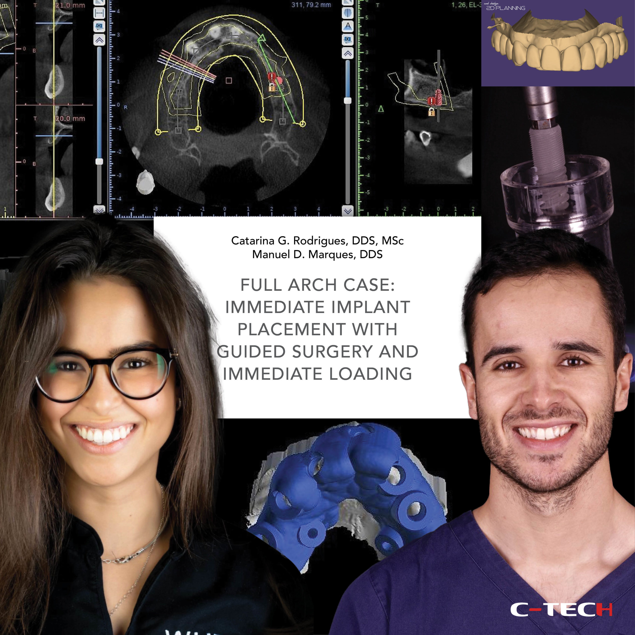
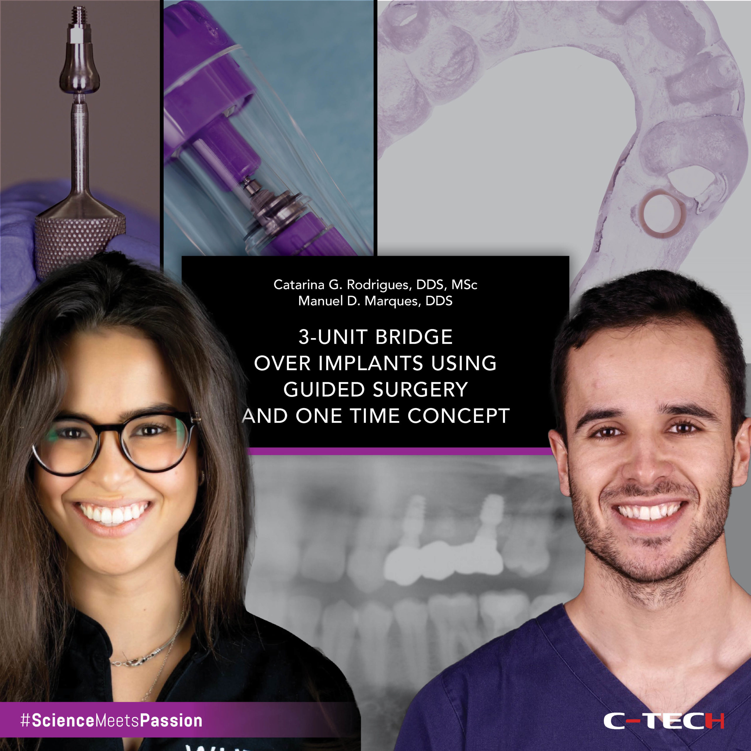
Catarina G. Rodrigues, DDS, MSc – Manuel D. Marques, DDS
A 66-year-old man presented to a private practice seeking a fixed restoration to replace his terminal dentition. The intraoral and radiographic examination revealed partially edentulous arches, the presence of periapical infections and extensive carious lesions on several teeth, significant attachment loss, and tooth mobility (Fig.1-3).
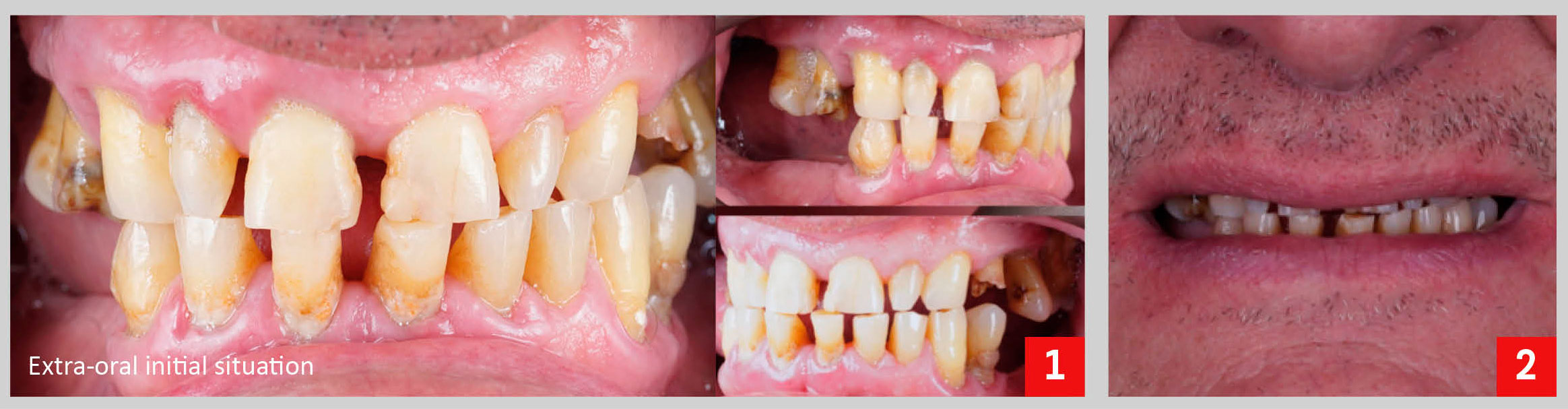
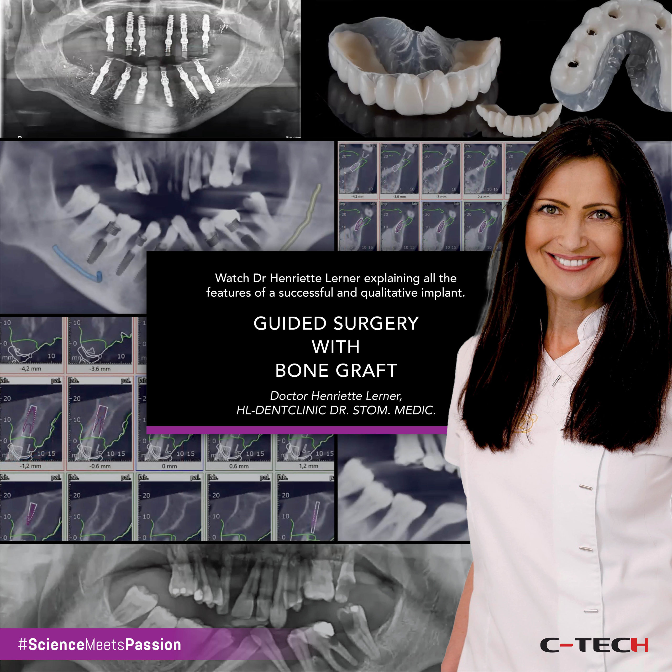
Both aesthetics and function were compromised. The vertical dimension of occlusion was reduced due to loss of posterior support and excessive wear. Following a proper diagnosis, the treatment plan proposed was the extraction of all the remaining teeth, placement of five implants in the maxilla and six implants in the mandible, immediate loading of the implants, and – as final restorations – full arch screw-retained prosthesis (Fig.4).

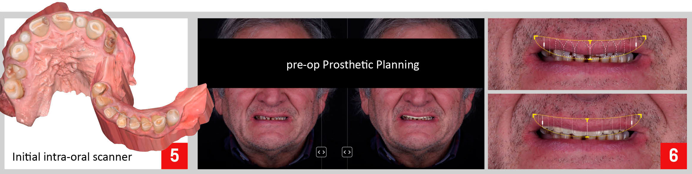
In order to perform proper pre-surgical planning of the case, initial records of the patient were obtained: intra and extra-oral photographs, digital impressions, and CBCT. A 2D facially driven digital smile design was made to aid in the planning of the position and dimension of the teeth for the future interim prosthesis.
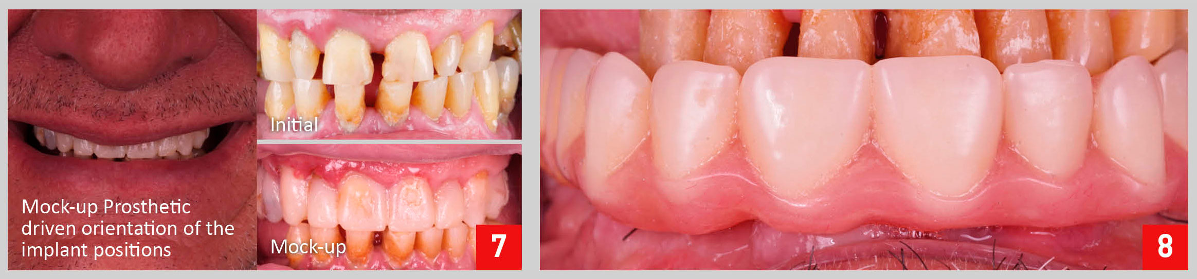
Then, using a specific 3D CAD software, a digital diagnostic wax-up was generated and 3D printed. A silicone index was obtained from the 3D printed model and filled with bis-acryl resin to produce trial restorations and evaluate the 2D smile planning on the patient’s mouth (Fig.5-7).
The approved try-in was then scanned and superimposed with the preoperative intra-oral scan and CBCT to digitally plan the implant surgery. Once the future implant positions were defined, they were translated into the design of the surgical templates (Fig.9-10).
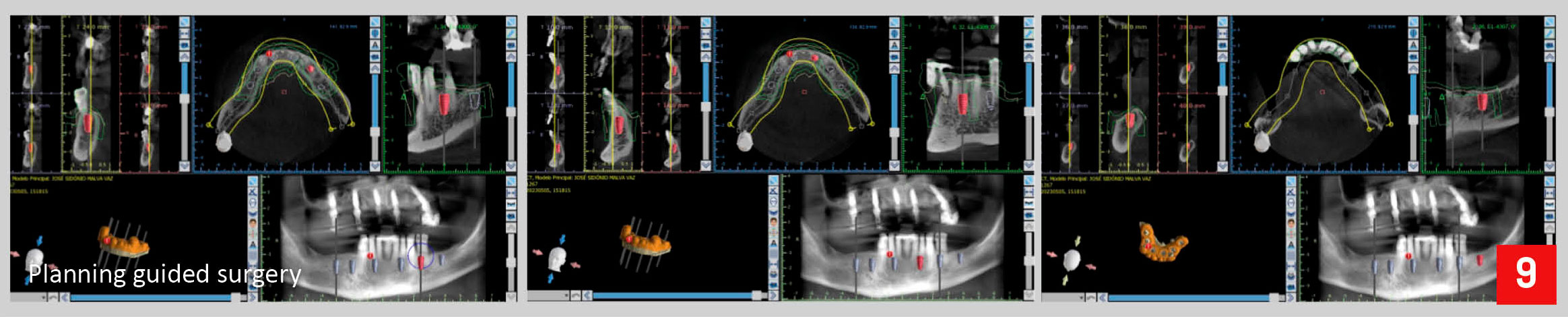
The prosthetic abutments were also planned in the same software as the implants.
Due to the patient’s systemic conditions, was decided to perform first the upper surgery and 3 months after the lower one. In the upper arch, after extraction of all maxillary hopeless teeth, implant sites were prepared through the guide according to a specific drilling protocol, and using C-Tech guided surgery kit, followed by implant placement.
All implants were torqued with at least 45Ncm to ensure enough primary stability for immediate loading. After implant placement, multi-unit abutments were inserted and torqued in place with 25 Ncm. A full arch provisional screw restoration was delivered the same day (Fig.8).
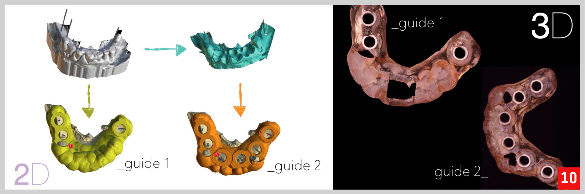
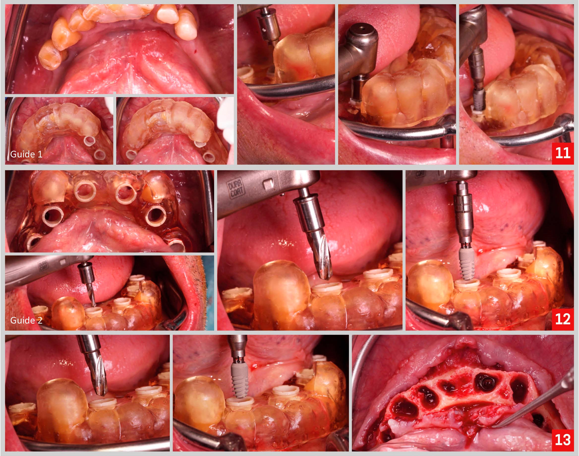
The implant surgery in the mandible was carried out 3 months after. To improve the precision of lower implant surgery, two surgical guides were used to place the implants.
The first guide was placed before any extraction and used to place implants on the molars region and right premolar region. Then, the first guide was removed, all teeth but the canines were extracted, and the second guide was seated and used to place implants on the anterior and left premolar regions (Fig. 11-13).
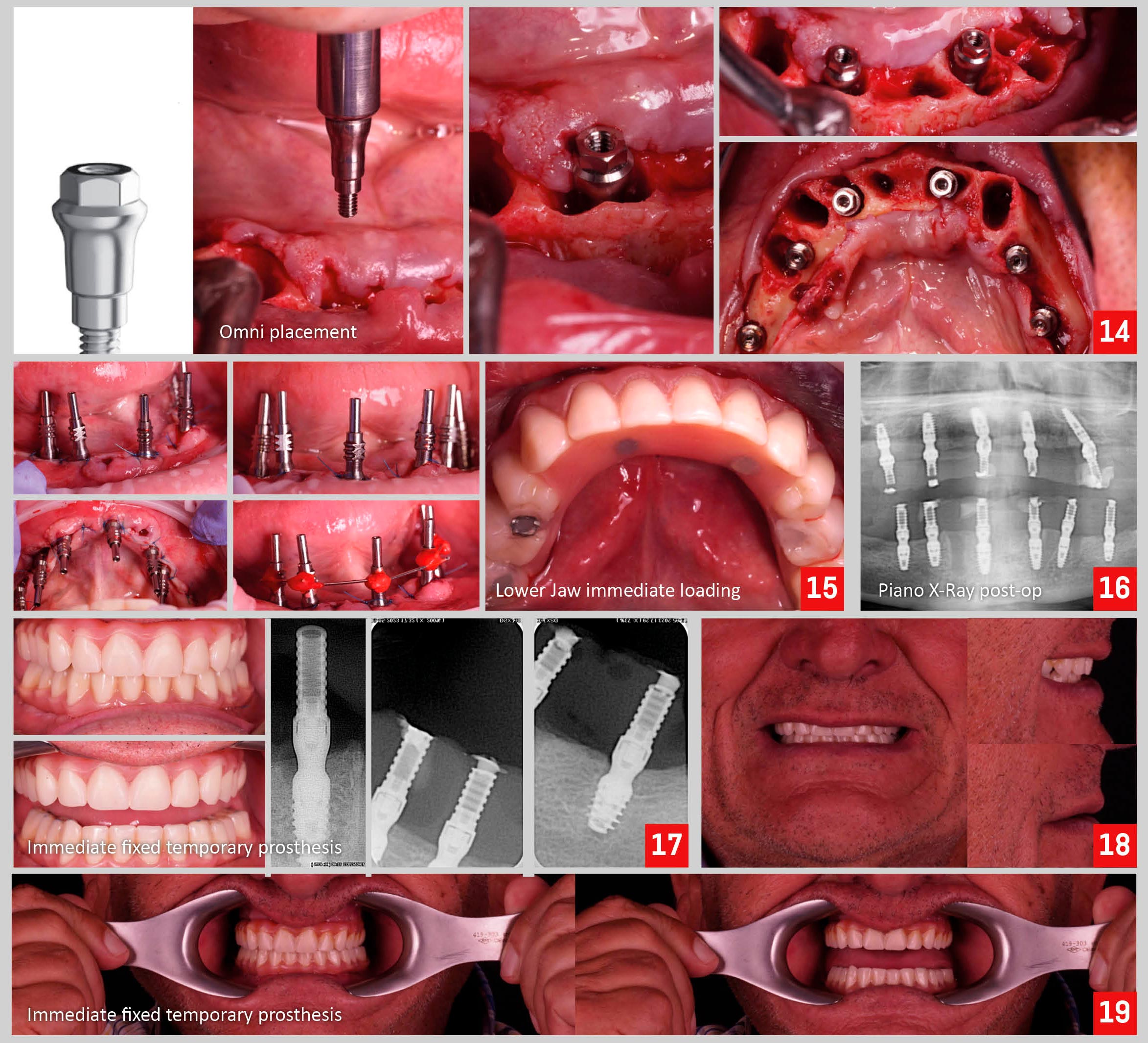
After implant placement, Omni system abutments were inserted and torqued in place with 25 Ncm. A full arch provisional screw restoration was delivered the same day. Post-op periapical x-rays and a panoramic x-ray were taken to ensure proper adaptation of the interim prosthesis to the implant abutments. Intra and extra-oral pictures of the immediate prosthesis were obtained (Fig. 14-19).
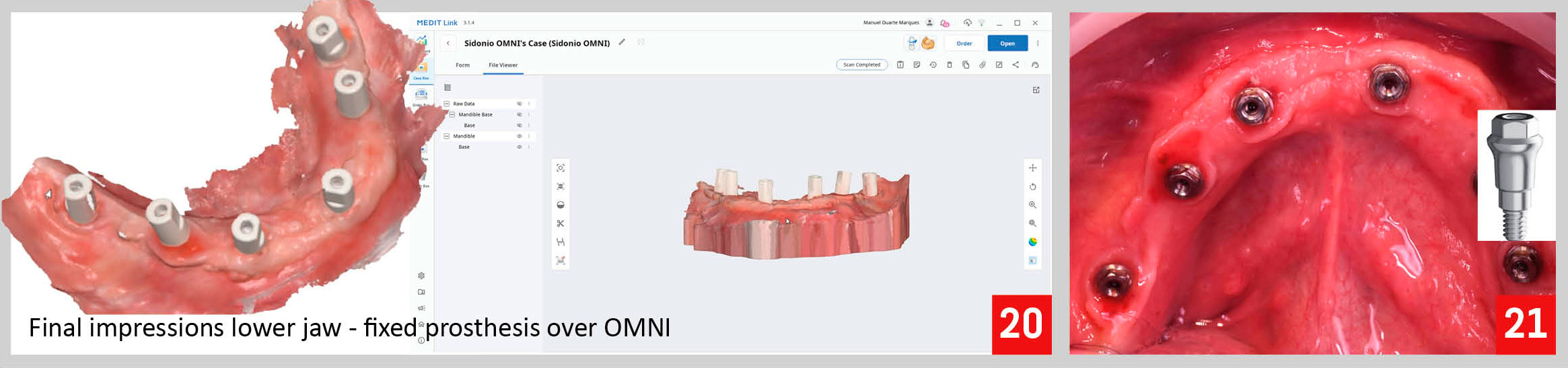
Three months after the lower, and six months after the upper surgery and immediate loading, we proceed with final impressions. In an occlusal view, after removing the lower fixed provisional prosthesis, we can appreciate a healthy appearance of the soft tissue (Fig. 20,21).
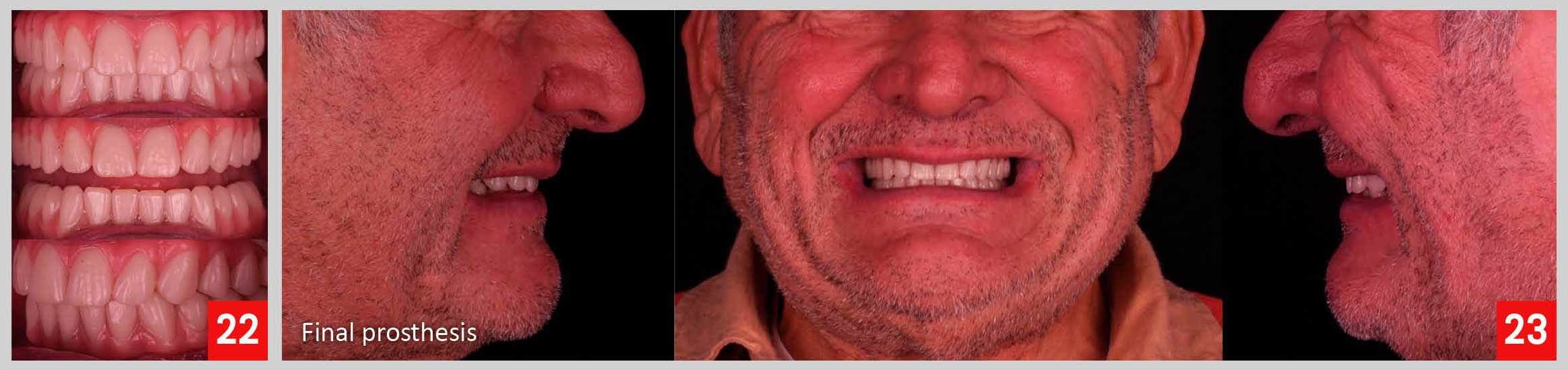
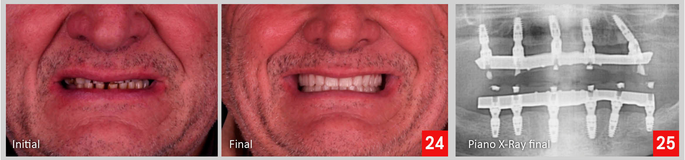
Finally, we deliver a full arch screw retained fixed prosthesis over multi-unit abutments in the upper arch, and a full arch screw retained fixed prosthesis over Omni system abutments in the lower arch (Fig. 22-25).