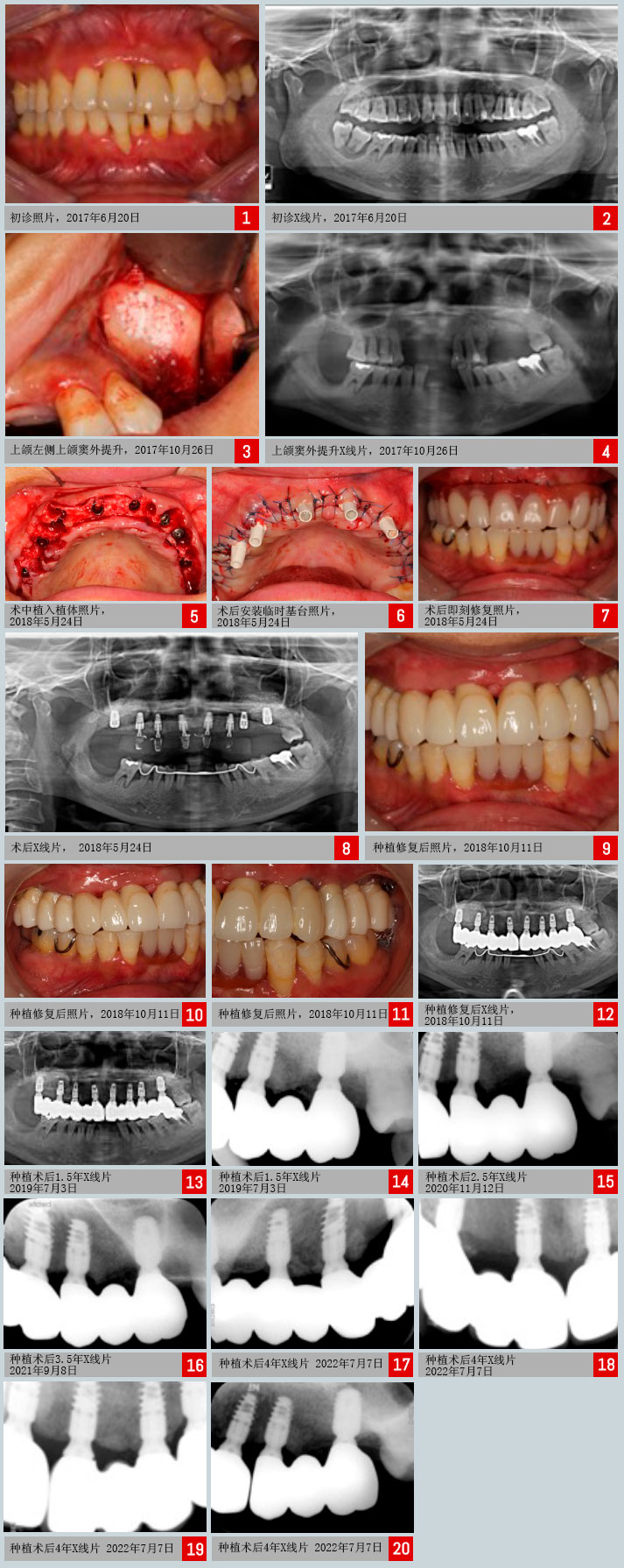
info@c-tech-implant.com +39 051 66 61 817 + 49 721 60 95 32 38

    • Italiano
    • English
    • Deutsch
    • Español
    • Français
    • Русский
    • 中文 (中国)
    • Türkçe
    • Ελληνικά
    • Magyar
    • Српски језик
    • Lietuviškai
    • Bahasa Indonesia
    • Slovenčina

Slide Mission, Internazionale, Garanzia, Certificazione, prodotti Compatibili

EL / Esthetic Line

病例报告表

Written by feRKzzCTo0 on . Posted in EL – Esthetic Line, Peng Dong, 传统种植学, 全弓, 即刻负荷, 臨床病例, 骨再生

Doctor Peng Dong, the expert of tooth implantation in Peking University International Hospital and president of Beijing Hedu Stomatological Clinic Co., Ltd

Medical Case Report

性别/年龄: 女/ 37岁  
初诊: 09/01/2014 植牙: 24/05/2018
辅助措施: 26/10/2017 最终修复: 11/10/2018
第1次复诊: 18/10/2018  
患者主诉: 上颌牙齿松动,影响进食求诊。
特殊事项: 即刻种植+即刻负重+上颌窦外提升术+GBR

病例概要

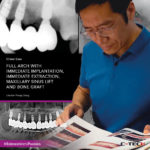
上颌牙齿牙槽骨吸收明显,根尖周大面积暗影,根吸收达根长1/3~1/2,诊断侵袭性牙周炎,经上颌窦外提升术后,拔除患牙,即刻植入C-Tech种植体,型号:EL-3509、EL-4309、EL-5109,共8颗,后期氧化锆烤瓷冠桥修复。

治疗计划

上颌牙槽骨宽度理想,骨高度不足,双侧上颌窦外提升术后2个月,拔除患牙,即刻植入C-Tech种植体EL-3509、EL-4309、EL-5109,共8颗,同期植骨后,植体支持的固定临时义齿修复。

治疗内容

1、常规消毒铺巾。上颌术区局部浸润麻醉。
2、行右上7远中斜行切口+右上7牙槽嵴顶近远中向切口+右上6543沟内切口+右上2~左上1牙槽嵴顶近远中向切+左上234沟内切口+左上5~7牙槽嵴顶近远中向切口,左上8沟内切口,翻瓣,暴露骨面,钳除右上6543和左上234清创,咬骨钳+大球钻降低平整骨面。
3、右上1定点,级差备洞+骨挤压至3.0*11mm,植入C-Tech种植体EL-3509+EL-4504P peek基台,扭力45N,边缘骨高度M1.5D1.5B1.5L1。右上3和左上1、3 定点,级差备洞+骨挤压至3.0*1 1mm,植入C-Tech种植体EL-3509+EL-4504P peek基台,左上1扭力45N,边缘骨高度M 2.5D2B1.5L2。左上3扭力45N,边缘骨高度M 1.5D1.5B-1.5L1.5。右上3扭力45N,边缘骨高度M 2D1.5B2L1.5。右上4定点,级差备洞至3.8*11mm,植入C-Tech种植体EL-4309+EL-4504P peek基台,扭力45N,边缘骨高度M1.5D2B1.5L1.5。左上4定点,级差备洞至3.8*11mm,植入C-Tech种植体EL-4309,扭力10N,边缘骨高度M1.5D-8B1.5L1.5。左右上6定点,级差备洞至3.8*11mm,各植入C-Tech种植体EL-5109+覆盖螺丝,左上6扭力25N,边缘骨高度M1D1B1.5L0.5。右上6扭力45N,边缘骨高度M1.5D1.5B1.5L1.5。将骨粉Bio-Oss分别置于右上3、1和左上1、3、4骨缺损出及唇侧骨板处,覆盖Bio-Gide于植骨处,及右上6拔牙处,拉拢并间断缝合关闭创口。
4、利用右上4、3、1和左上1、3放置临时基台,制作上颌即刻义齿,调颌,抛光。
5、九个月后取模,更换修复基台,粘接固定,氧化锆烤瓷冠桥永久修复。


Insertion of six EL implants in guided surgery

Written by feRKzzCTo0 on . Posted in Alexandre Minis, EL – Esthetic Line, GS – Guided Surgery, 导引式植牙, 臨床病例

Dr. Alexandre Minis, France

The patient is a 52-year-old male with no medical history.
Chronic periodontitis is stabilized.
Former smoker who quit following periodontal treatment.
Teeth 26, 16 and 17 were extracted. Considering that the available bone height was low, a bilateral sinus lift was performed by lateral approach.

Webinar | EL Esthetic Line: What are the ingredients of a great implant?

Written by feRKzzCTo0 on . Posted in EL – Esthetic Line, Henriette Lerner, 传统种植学, 臨床病例

Watch Dr Henriette Lerner explaining all the features of a successful and qualitative implant.

I saw you have breathing plant systems in the pipeline and you learn a lot of systems, and probably you already have in your mind some main features that the implants need to have.
In order to make your treatment easier and more predictable in your procedures.
You probably already know that an implant has to have a high bone implant contact surface and the designs of the implants are all made in a way that increases this surface, because this is giving you a long-term stability of the surface of the osteointegration.

Insertion of two EL implants on the upper jaw in guided surgery with immediate loading

Written by feRKzzCTo0 on . Posted in EL – Esthetic Line, GS – Guided Surgery, Luigi Ciacci, 即刻负荷, 导引式植牙, 臨床病例

Dr. Luigi Ciacci, Odt. Andrea Sessa

Introduction:
Modern dentistry depends on comprehensive diagnostics and careful planning to achieve the desired result and meet the expectations of both the patient and the dentist.
Digital planning and guided surgery based on three-dimensional X-ray data and digitized intraoral recording are of great help. They provide valuable information and allow for accurate backward planning to optimize the implant-prosthetic outcome, making the restoration more predictable in terms of function, aesthetics and biology.

Guided Implant on a Tricky Crest – clinical case

Written by feRKzzCTo0 on . Posted in Alexandre Minis, EL – Esthetic Line, GS – Guided Surgery, 导引式植牙, 臨床病例

Dott. Alexandre Minis, France

Patient is a 56 years old male without a previous medical history. Tooth # 36 has been missing for several year.
Despite a satisfactory bone height on the panoramic, the CBCT reveals a thin bone at the level of the crest which does not allow the available height to be exploited.
In order to obtain an ideal prosthetic axis and to use the available bone to the maximum, the surgery is performed using a surgical guide, design on 3Shape Implant Studio, a classic dentition supported guide, 3D printed on a Phrozen Shuffle printer.

Extraction of a Fractured Upper Left Second Premolar, Conventional Implant Placement, with Bone Grafting and Immediate Loading of a Provisional. Final restoration with digital occlusal analysis using T-Scan. – A Case Report

Written by feRKzzCTo0 on . Posted in EL – Esthetic Line, Fabrizia Luongo, 传统种植学, 即刻负荷, 臨床病例

Dr Fabrizia Luongo, DDS, MS, Periodontist, Rome, Italy

Introduction
The evolution of digital dentistry and the development of a digital workflow has concentrated on digital planning with the use of Cone Beam CT scanning as well as using digital restorative tools to combine DICOM (Digital Imaging Communication in Medicine) and .stl (stereolithography) files to virtually plan, place and restore implants before using this plan to treat patients. The resulting benefits are reduced chair time, high precision and predictable aesthetic results often with immediate fixed provisional restorations available at time of surgery and corresponding high levels of patient satisfaction.
Intra-oral scanning to create digital ‘virtual impressions’ is also becoming more prevalent with the information being stored in the .stl file format. This information can be utilised by appropriate CAD/CAM (computer-aided design and computer aided manufacturing) software to design and manufacture a dental restoration (either by milling or 3D printing).
One area that is sometimes overlooked is the use of digital technology in occlusal analysis and adjustment of the restored dental implant. The following case study examines the occlusal management of a conventionally placed implant.

Extraction of endodontically failed pre-molar and immediate placement of a C-Tech Esthetic Line implant utilising a Guided Surgery Technique – a Case Report

Written by feRKzzCTo0 on . Posted in EL / Esthetic Line, EL – Esthetic Line, Fabrizia Luongo, GS – Guided Surgery, 导引式植牙, 臨床病例

Dr Fabrizia Luongo, DDS, MS, Periodontist, Rome, Italy

Introduction
The use of Cone Beam Computerised Tomography (CBCT) scanning is becoming more commonplace in today’s implant dentistry. These scans combined with increasingly sophisticated software technology has led to greater accuracy in digital planning and guided surgery and is gradually being adopted across general practice. Furthermore, the restorative planning can be digitised and combined with the CBCT data in appropriate planning software.
In conjunction with these technology advances, dental implant design has evolved to incorporate the latest concepts of biomechanical design, which include sophisticated thread profiling, platform switching and a Morse locking taper on the implant/abutment interface.
The following case details brings these advances in implants and digital technologies together.

Placing a C-Tech Esthetic Line Implant Utilising a Navigated Surgery Approach – A Case Report

Written by feRKzzCTo0 on . Posted in EL / Esthetic Line, EL – Esthetic Line, Fabrizia Luongo, GS – Guided Surgery, 导引式植牙, 臨床病例

Dr Fabrizia Luongo, DDS, MS, Periodontist, Rome, Italy

Introduction
There has been a considerable evolution in implant dentistry over recent years that has seen the design of dental implants adopt sophisticated thread profiles which leads to a better primary stability. Simultaneously these new geometries aid cortical bone maintenance through to platform-switching capabilities again designed to minimise bone loss. When combined with improved implant-prosthetic connections such as a Morse-locking conical connection it contributes to a good long-term prognosis and an aesthetic outcome.
Concurrent with this evolution in implant design have been substantial advances in digital technologies across the field of dentistry. These digital advances include Cone-beam CT scanning combined with appropriate 3D planning software, navigated surgery technology, 3D intra-oral scanning to create a ‘virtual impression’ and 3D printing technologies. These digital technologies can minimise the number of appointments found in a conventional treatment protocol as well as enabling greater accuracy and will be considered in this case study.

Immediate loading of a morse locking conical implant with C-TECH EL; Case report

Written by feRKzzCTo0 on . Posted in EL / Esthetic Line, EL – Esthetic Line, SD-MB – Monoblock - Small Diameter, 传统种植学, 即刻负荷, 微种植体, 臨床病例

A 55 year old female patient was referred to our office for a full mouth rehabilitation. Upon clinical examination and a CBCT, we have decided to implant 4 EL C-Tech conical Morse tapered connection implants in the upper jaw for a bar retained over denture and due to financial considerations; was to perform extraction of all remaining lower teeth and immediate post extraction implantation of SD C-Tech mini dental implants. A full muco-periosteal flap was released, teeth were extracted, an alveotomy was performed to achieve an optimal bone platform for the SD mini dental implants and to obtain a bigger vertical dimension for the overdenture!

Prefooter CN