• Italiano
    • English
    • Deutsch
    • Español
    • Français
    • Русский
    • 中文 (中国)
    • Türkçe
    • Ελληνικά
    • Magyar
    • Српски језик
    • Lietuviškai
    • Bahasa Indonesia
    • Slovenčina

Slide Mission, Internazionale, Garanzia, Certificazione, prodotti Compatibili

EL / Esthetic Line

Advantages of guided surgery, predictability in small spaces

Written by feRKzzCTo0 on . Posted in EL – Esthetic Line, GS – Guided Surgery, Henriette Lerner, Вођена Хирургија, Клинички случајеви

by Doctor Henriette Lerner, HL-DENTCLINIC DR. STOM. MEDIC.

In this case, the patient has a genesis of the two upper lateral incisors.
Having created the space with the help of the orthodontic specialist, we first proceed with a Digital Smile Design and then with the digital planning of the two implants in comparison with the new aesthetics.

Guided surgery in the jaw and mandible

Written by feRKzzCTo0 on . Posted in EL – Esthetic Line, GS – Guided Surgery, Henriette Lerner, Вођена Хирургија, Клинички случајеви

by Doctor Henriette Lerner, HL-DENTCLINIC DR. STOM. MEDIC.

In this case, it is necessary to use all the digital tools available – detection of joint movements, digital smile design and guided surgery, with the aim of having predictability and accuracy. For the future aesthetic part and function, the evaluation took place before the surgery.

Guided surgery with bone graft

Written by feRKzzCTo0 on . Posted in EL – Esthetic Line, GS – Guided Surgery, Henriette Lerner, Вођена Хирургија, Клинички случајеви

by Doctor Henriette Lerner, HL-DENTCLINIC DR. STOM. MEDIC.

In this case it is shown how it is still possible to work in guided surgery and consequently perform bone grafts where necessary.
The advantage in a difficult case like this is to design the dental implants with a surgical guide, in order to maintain their correct positioning.
Finally, the aesthetic project was evaluated prior to surgery.

One Time Clever Concept: Non-removal of immediate abutments in a single crown and 3-unit bridge over implant

Written by feRKzzCTo0 on . Posted in Catarina G. Rodrigues, EL – Esthetic Line, Manuel D. Marques, ONE TIME Clever Concept, Клинички случајеви, Конвенционална Имплантологија

Catarina G. Rodrigues, DDS, MSc – Manuel D. Marques, DDS

A 67-year-old female patient presented to a private dental office with pain on the first quadrant. The clinical and radiographic examination revealed extensive caries and a history of root canal therapy failure on teeth #1.3 and 1.5. Also, absence of teeth #1.4 and #1.6 (Fig. 2-4).

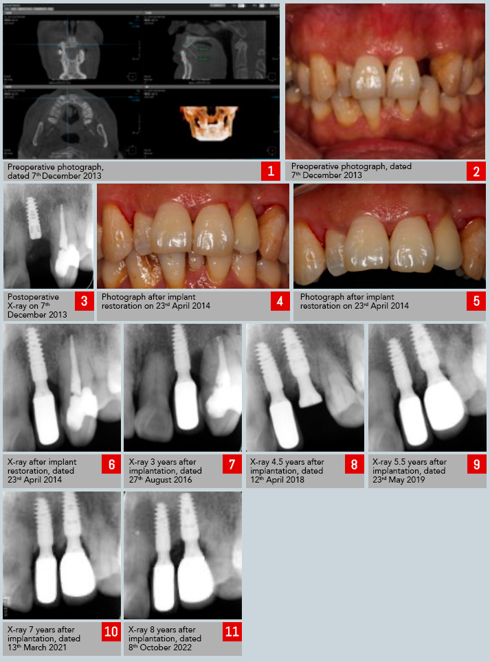
2 implants restoration in the upper jaw, 8-year follow-up

Written by feRKzzCTo0 on . Posted in EL – Esthetic Line, Peng Dong, Клинички случајеви, Конвенционална Имплантологија

Doctor Peng Dong, the expert of tooth implantation in Peking University International Hospital and president of Beijing Hedu Stomatological Clinic Co., Ltd

Medical Case Report

Sex / Age: Male/55  
First Visit: 07/12/2013 Implant Surgery: 07/12/2013
Others Surgery (GBR…): Final Restoration: 23/04/2014
First Recall: 30/04/2014  
Chief Complaint: Missing an anterior tooth in the upper left region, the patient seeks fixed restoration.
Consideration(s):

CASE OUTLINE

The upper left tooth 2 is missing. It will undergo restoration using a C-Tech implant (Model: EL-3511). A subsequent application of an oxidized zirconia porcelain crown is planned for the final restoration.

TREATMENT PLAN

The width of the alveolar bone in the region of the upper left tooth 2 measures 5.7mm, and the height is 14.02mm, which is deemed to be ideal. The proposed approach is to implant a C-Tech implant (Model: EL-3511) in this area. The second stage of the surgery is scheduled for three months later.

TREATMENT

1、Perform standard disinfection and drape the area with cloths.
2、Administer local anesthesia to the upper left tooth 2, ensuring minimal discomfort. Create a horizontal incision along the crest of the alveolar ridge, extending from the near to far and central aspects. Carefully reflect the gingiva to expose the underlying bone surface.
3、Precisely position the C-Tech implant (Model: EL-3511) in the location of the upper left tooth 2. Gradually prepare a tapered borehole measuring 3.0x13mm to accommodate the implant, ensuring a precise fit. The implant is integrated with the healing abutment (Model: EL-5506HT) and anchored with a torque of 10N, achieving a marginal bone height of M3D4 B1.5L2. Securely suture the area.
4、After 4 months, take an impression and replace the restorative abutment with Model: EL-4504F. Permanently fix it with adhesive fixation, and apply an oxidized zirconia porcelain crown for long-lasting restoration.


Insertion of six EL implants in guided surgery

Written by feRKzzCTo0 on . Posted in Alexandre Minis, EL – Esthetic Line, GS – Guided Surgery, Вођена Хирургија, Клинички случајеви

Dr. Alexandre Minis, France

The patient is a 52-year-old male with no medical history.
Chronic periodontitis is stabilized.
Former smoker who quit following periodontal treatment.
Teeth 26, 16 and 17 were extracted. Considering that the available bone height was low, a bilateral sinus lift was performed by lateral approach.

Webinar | EL Esthetic Line: What are the ingredients of a great implant?

Written by feRKzzCTo0 on . Posted in EL – Esthetic Line, Henriette Lerner, Клинички случајеви, Конвенционална Имплантологија

Watch Dr Henriette Lerner explaining all the features of a successful and qualitative implant.

I saw you have breathing plant systems in the pipeline and you learn a lot of systems, and probably you already have in your mind some main features that the implants need to have.
In order to make your treatment easier and more predictable in your procedures.
You probably already know that an implant has to have a high bone implant contact surface and the designs of the implants are all made in a way that increases this surface, because this is giving you a long-term stability of the surface of the osteointegration.

Insertion of two EL implants on the upper jaw in guided surgery with immediate loading

Written by feRKzzCTo0 on . Posted in EL – Esthetic Line, GS – Guided Surgery, Luigi Ciacci, Вођена Хирургија, Клинички случајеви

Dr. Luigi Ciacci, Odt. Andrea Sessa

Introduction:
Modern dentistry depends on comprehensive diagnostics and careful planning to achieve the desired result and meet the expectations of both the patient and the dentist.
Digital planning and guided surgery based on three-dimensional X-ray data and digitized intraoral recording are of great help. They provide valuable information and allow for accurate backward planning to optimize the implant-prosthetic outcome, making the restoration more predictable in terms of function, aesthetics and biology.

Guided Implant on a Tricky Crest – clinical case

Written by feRKzzCTo0 on . Posted in Alexandre Minis, EL – Esthetic Line, GS – Guided Surgery, Вођена Хирургија, Клинички случајеви

Dott. Alexandre Minis, France

Patient is a 56 years old male without a previous medical history. Tooth # 36 has been missing for several year.
Despite a satisfactory bone height on the panoramic, the CBCT reveals a thin bone at the level of the crest which does not allow the available height to be exploited.
In order to obtain an ideal prosthetic axis and to use the available bone to the maximum, the surgery is performed using a surgical guide, design on 3Shape Implant Studio, a classic dentition supported guide, 3D printed on a Phrozen Shuffle printer.

Complete-arch fixed reconstruction by means of guided surgery and immediate loading: a retrospective clinical study on 12 patients with 1 year of follow-up

Written by feRKzzCTo0 on . Posted in El – Esthetic Line, Gs – Guided Surgery, Henriette Lerner, Nd – Narrow Diameter, Robert Sader, Scientific Publications, Shahram Ghanaati, Uli Hauschild, Вођена Хирургија, Конвенционална Имплантологија

Henriette Lerner, Uli Hauschild, Robert Sader & Shahram Ghanaati

Abstract
Guided implant surgery is considered as a safe and minimally invasive flapless procedure. However, flapless guided surgery, implant placement in post-extraction sockets and immediate loading of complete-arch fixed reconstructions without artificial gum are still not throughly evaluated. The aim of the present retrospective clinical study was to document the survival and success of complete-arch fixed reconstructions without artificial gum, obtained by means of guided surgery and immediate loading of implants placed also in fresh extraction sockets.

Rehabilitation of the jaw by means of immediate implantation and immediate restoration Digital workflow

Written by feRKzzCTo0 on . Posted in Catarina G. Rodrigues, El – Esthetic Line, Gs – Guided Surgery, Manuel D. Marques, Mua, Scientific Publications, Вођена Хирургија, Конвенционална Имплантологија

Magazine PIP • Practical Implantology and Implant Prosthetics • August 2022 | Issue 4′

Rehabilitation of the jaw by means of immediate implantation and immediate restoration Digital workflow

Catarina G. Rodrigues, DDS, MSc – Manuel D. Marques, DDS – Raquel Bandeira, CDT

The success of any complex dental treatment depends on proper diagnostics and treatment planning. Digital treatment planning, prosthetic backward planning and guided implant surgery can help improve success rates and predictability of complex rehabilitations. The 3D information about the bone anatomy obtained by means of digital volume tomography (DVT) of the patient form the basis for successful treatment planning.
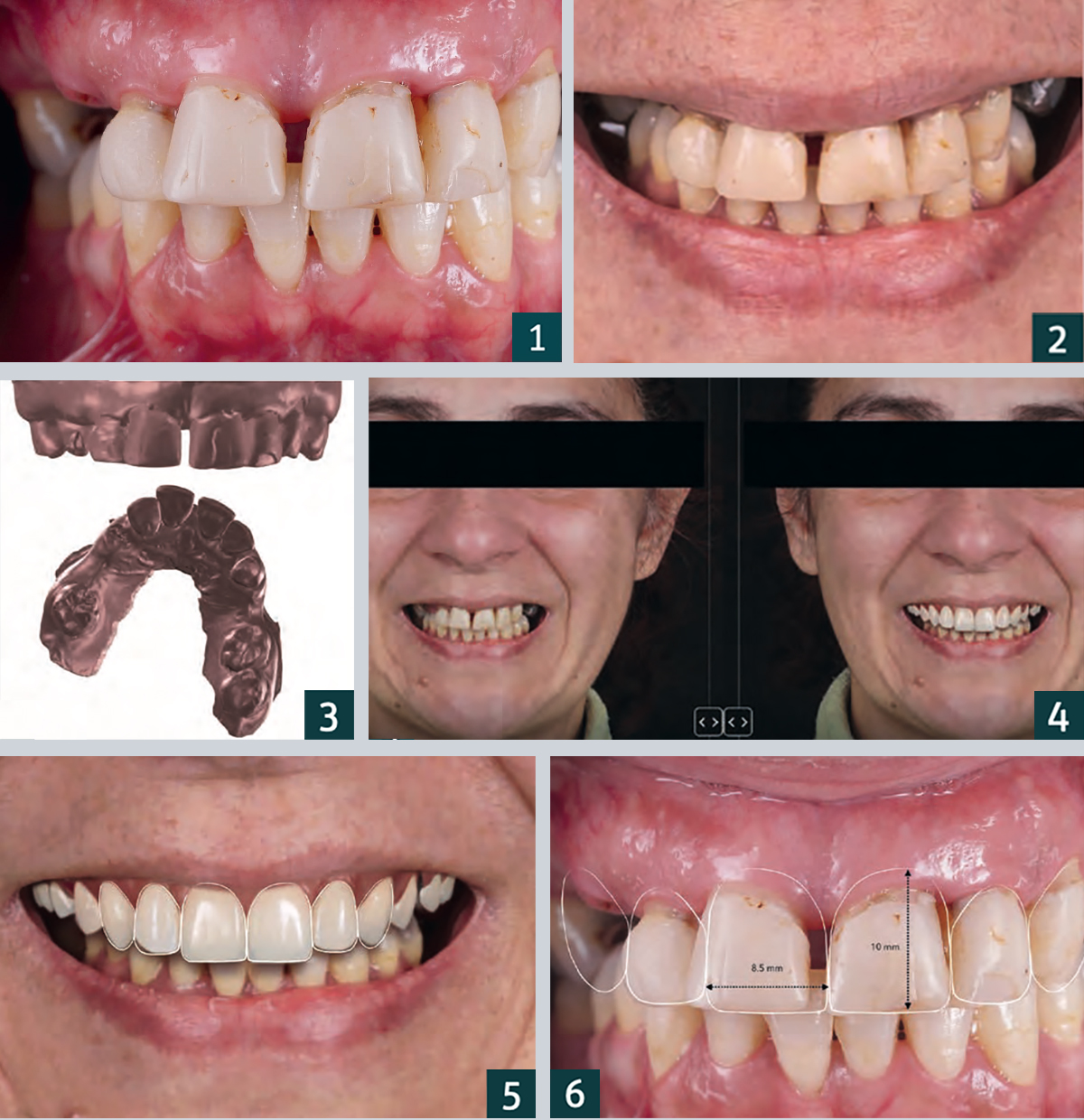
1. Frontal view with centric occlusion preoperative.
2. Preoperative close-up of the patient’s smile.
3. Image data for the preoperative intraoral scan.
4. Virtual simulation of the prosthetic reconstruction using 2D CAD software (Smile Cloud Biometrics; ADN3D Bioetch SRL).
5. Virtual planning of the shape and position of the anterior teeth according to the measurement criteria ..
6. … the red-and-white aesthetic, planned with the 2D Facial Smile design.

PROCEDURE
The DVT data is imported and processed by means of appropriate software in order to plan the implant positions. The planning data is then used to virtually design the drilling template for guided implant placement, which is then 3D printed and used for optimal positioning of the implants. The use of 2D dental CAD software enables the development of a face-related digital smile design. This two-dimensional data can then be imported into 3D CAD software to design 3D mock ups and immediate temporary restorations.

Advantages of immediate implantation
Immediate implantation of implant-supported prostheses is a predictable treatment option in the restoration of an edentulous jaw, as it can contribute to the preservation of the peri-implant soft and hard tissue and enable rapid patient assistance from an aesthetic and functional point of view. Important advantages of using digital technologies for the planning and execution of implant placement as well as for interim restoration are higher accuracy and precision in the fabrication of the overall rehabilitation, low invasiveness and optimal soft tissue shaping by the temporary prosthesis.
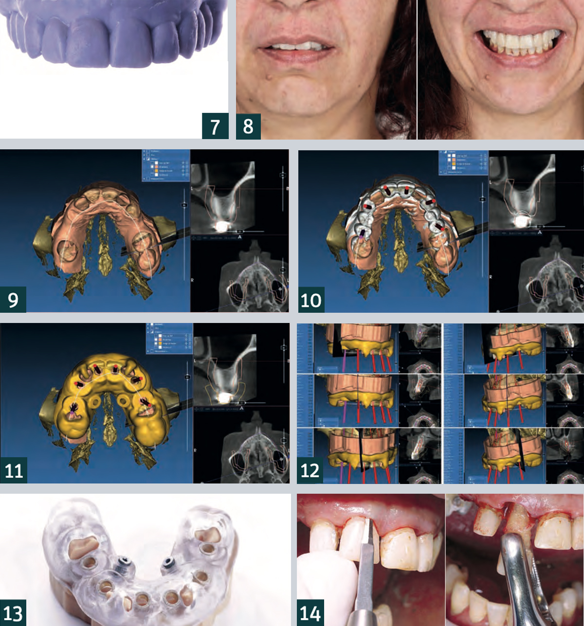

7. Frontal view of the 3D-printed model created from the diagnostic wax-up.
8. Frontal view of the preoperative mock up after transfer to the patient, checking the planning of the 2D facial smile design.
9. Superimposition of the STL data of the preoperative intraoral scan with the DVT data in the planning software (RealGUIDE, 3DIEMME).
10. Digital planning of the implants based on the anatomical situation and the previously planned prosthetic restoration.
11. Once the final implant positions were determined, they were transferred to the surgical template design.
12. Planning of the implant positions.
13. Adjustment of the drilling template on the model.
14. Gentle extraction of the teeth not worth preserving in order to preserve the hard and soft tissue dimensions.

Patient case
The 40-year-old patient presented to our dental practice complaining in particular of difficulty eating and worsened aesthetics in the maxillary anterior region (Figs. 1, 2). The clinical and radiographic examination revealed a bilateral interdental gap in the maxillary posterior region. In addition to a fracture of tooth 13 at gingival level, insufficient amalgam and composite fillings as well as carious defects were diagnosed. There were also increased probing depths and generalised, radiographically visible, horizontal bone loss. After comprehensive diagnostics, it was decided, in consultation with the patient, to extract all of the upper anterior and posterior teeth, and proceed with immediate implantation of a total of eight implants and immediate provisional restoration with screw-retained dentures made of polymethyl methacrylate (PMMA). Intraoral scans (Fig. 3), a DVT and intraoral and extraoral photos (Fig. 4) were taken as part of preoperative diagnosis. In addition, a 2D digital facial smile design was created to enable planning of the position as well as the shape and size of the teeth for the future interim prosthesis (Figs. 5, 6). A digital diagnostic wax-up was then created using 3D CAD software and transferred to a corresponding model using 3D printing (Fig. 7).

Based on the model, a silicone index was created to enable the transfer of the acrylic resin trial restorations to the patient situation and 2D smile planning was undertaken for the patient’s mouth by means of a mock-up (Fig. 8). After adjusting and matching the shape and aesthetics of the anterior teeth, all three-dimensional data (DVT, preoperative intraoral scan and 3D mock-up) were imported into software (Fig. 9) and used to plan the implant position and fabricate the surgical guide (Figs. 10-12). In the patient case under discussion, a mostly fully-navigated implant placement was planned. Special guide sleeves adapted to the implant system were therefore integrated into the surgical template (Fig. 13). The surgical guide was stabilised with tooth support on teeth 16, 12, 22, 26 and 27 and with two palatal bone screws for which two screw channels had been integrated into the surgical guide. Implant preparation was initially fully navigated in the extraction sockets of teeth 13, 11, 21, 23 without opening and in region 15 and 25 with the formation of a mucoperiosteal flap.

After insertion of the six standard implants in the anterior region, the surgical template was removed, followed by extraction of teeth 16, 12, 22, 26 and 27 (Figs. 14-25). The placement of the two short implants was carried out freehand in region 16 and 26 after removal of the posterior teeth. All implant preparations were carried out according to the manufacturer’s recommended drilling protocol. The provisional prosthesis (Fig. 26) was screwed onto the six anterior implants via abutments and immediately loaded (Fig. 27). A postoperative X-ray was taken (Fig. 28). The patient is very satisfied with her new restoration, both aesthetically and functionally (Figs. 29-31).
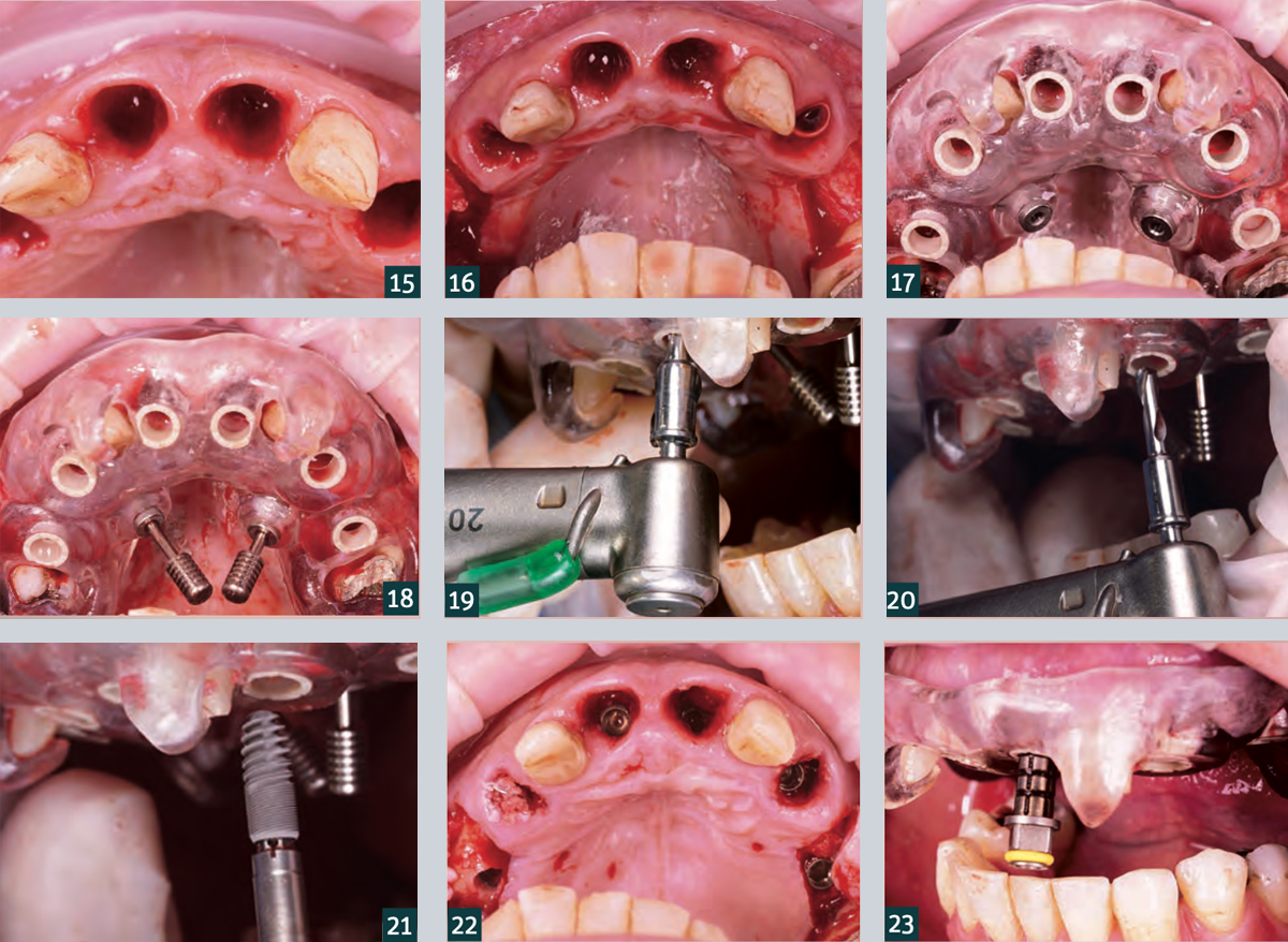
15. Teeth 12 and 22 were initially left in place to stabilise the surgical guide.
16. A full thickness flap was mobilised buccally in the premolar region of the quadrants to increase the soft tissue thickness.
17. Check the intraoral fit and stability of the surgical guide.
18. Additional stabilisation of the surgical guide via palatal fixation screws.
19. Template-guided implant drilling using the C-Guide drill..
20. … for C-Tech implants (C-Tech Implant, Bologna).
21. Template-guided implant placement (Esthetic Line implants, C-Tech Implants).
22. Occlusal view after placement of the six fully-navigated anterior implants.
23. Control: The markings on the transfer post correspond to the surgical guide sleeves.
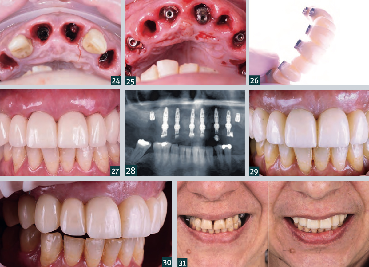

24. Occlusal view of the final implant position in apicocoronal direction (subcrestal positioning of the implants!).
25. Placement of the Multi Unit abutments.
26. Temporary denture made of PMMA.
27. Situation after insertion of the temporary denture with good postoperative soft tissue condition.
28. Radiograph showing postoperative check overview.
29. Intraoral situation one week after the procedure with…
30. … healing without complications and good soft tissue condition.
31. Comparison of the patient situation before (left) and one week after the intervention (right).
Small but powerful. High-quality range with C-Tech system implants.

Written by feRKzzCTo0 on . Posted in El – Esthetic Line, Nd – Narrow Diameter, Scientific Publications, Вођена Хирургија, Конвенционална Имплантологија, Мини Импланти, Регенерација Костију

Magazine PIP • Practical implantology and implant prosthetics • November 2019

Small but powerful. High-quality range with C-Tech system implants.

Full Smile, under the direction of business owner Lütfü Agic, well-known in the field of dental implantology, advises on a high-quality range with C-Tech system implants, biomaterials such as Bioteck, as well as dental and surgical instruments and tools from devemed. How can such a small unit survive in times of ever greater global players in the field of dentistry and what is the reason for a dentist to look for the partnership, asked pip and talked to Stefan Grümer, M.Sc., who runs together with colleagues a large referral practice on Theaterstrasse in Aachen.

pip: Don’t you kind of lose touch with international developments and trends with such a small partner like Full Smile?
Dr. Grümer: That may well be, regarding these international trends. Here, in the Theaterstraße in Aachen, we are a practice with an impressive range of modern services, especially in the field of restorative and aesthetic dentistry. From 3D diagnostics and computer-based functional diagnostics to laser dentistry, we use state-of-the-art and exceptionally gentle treatment methods that are comfortable for the patient. We are focusing strongly on training programs so that our range of services stays up to date with the latest state of science and research. But we do not have to follow any pseudo innovation. Much more important to us is a certain consistency with our commercial partners, because as well as between us and our patients, the cooperation between us and a medical device manufacturer is a matter of trust. It would bother me to have to adjust to a new contact person every few months, who knows neither me nor my treatment philosophy nor knows what my experiences are and what might actually enrich and interest me. Since 2013 I have been running the first university teaching practice in Germany in cooperation with the RWTH Aachen, an international academy with many foreign and extensive research projects – above all in the field of implantology and laser dentistry. Therefore, we are demanding a certain level of consulting expertise and a portfolio at eye level from our partners.

pip: What impressed you most about the C-Tech Implant System?
Dr. Grümer: As a referral practice we naturally work with different systems. Personally, I especially appreciate the C-Tech Implant System because of its system-related features. It is a fully engineered system that provides different implant diameters and lengths for all common indications – up to special designs such as one-piece mini-implants for narrow spaces, abutments or anchoring of dentures. At C-Tech we experience a very high primary stability and a very easy handling and thus a method which is absolutely suitable for everyday use. Furthermore, the aesthetic demands of our patients have increased significantly. With the EL – Esthetic Line – C-Tech offers a special design for these more demanding challenges. Refinements, like the platform switch, a generous apical threading, the morse taper connection and the concave aesthetic concept create a good stability with excellent shaping of the surrounding tissue. Despite a high prosthetic design freedom, the system is very easy to use, but the prosthetic connection is identical to the other implant diameters of the series. So you can get by with a straightforward instrumentation. I also like a whole new service of Full Smile, which I have never experienced before: As a dentist, I can search for any product via Full Smile and, if I found one, I will only pay the same list price as in the catalogue!

pip: What about service and accessibility – how can such a small company compete with companies that are quite different staffed?
Dr. Grümer: In fact, we sometimes ask ourselves the famous question of how often Mr. Agic has been cloned. He’s really frequently on the move and also visits us regularly to exchange ideas with us or to prepare ourselves for a new technique. However, we always reach someone in the office or receive a callback within a very short time – and also in such a short time our request is accepted, a delivery is initiated or a technical question is answered. In fact, with other companies I sometimes spent a lot more time in some telephone loop with automated speech before I can speak to a human being. With individual consultation, the reliable assurance of the desired delivery times and practical training, Full Smile is a professional partner for us in the field of dental implantology, periodontology and surgery.


Extraction of a Fractured Upper Left Second Premolar, Conventional Implant Placement, with Bone Grafting and Immediate Loading of a Provisional. Final restoration with digital occlusal analysis using T-Scan. – A Case Report

Written by feRKzzCTo0 on . Posted in EL – Esthetic Line, Fabrizia Luongo, Клинички случајеви, Конвенционална Имплантологија

Dr Fabrizia Luongo, DDS, MS, Periodontist, Rome, Italy

Introduction
The evolution of digital dentistry and the development of a digital workflow has concentrated on digital planning with the use of Cone Beam CT scanning as well as using digital restorative tools to combine DICOM (Digital Imaging Communication in Medicine) and .stl (stereolithography) files to virtually plan, place and restore implants before using this plan to treat patients. The resulting benefits are reduced chair time, high precision and predictable aesthetic results often with immediate fixed provisional restorations available at time of surgery and corresponding high levels of patient satisfaction.
Intra-oral scanning to create digital ‘virtual impressions’ is also becoming more prevalent with the information being stored in the .stl file format. This information can be utilised by appropriate CAD/CAM (computer-aided design and computer aided manufacturing) software to design and manufacture a dental restoration (either by milling or 3D printing).
One area that is sometimes overlooked is the use of digital technology in occlusal analysis and adjustment of the restored dental implant. The following case study examines the occlusal management of a conventionally placed implant.

Extraction of endodontically failed pre-molar and immediate placement of a C-Tech Esthetic Line implant utilising a Guided Surgery Technique – a Case Report

Written by feRKzzCTo0 on . Posted in EL / Esthetic Line, EL – Esthetic Line, Fabrizia Luongo, GS – Guided Surgery, Вођена Хирургија, Клинички случајеви

Dr Fabrizia Luongo, DDS, MS, Periodontist, Rome, Italy

Introduction
The use of Cone Beam Computerised Tomography (CBCT) scanning is becoming more commonplace in today’s implant dentistry. These scans combined with increasingly sophisticated software technology has led to greater accuracy in digital planning and guided surgery and is gradually being adopted across general practice. Furthermore, the restorative planning can be digitised and combined with the CBCT data in appropriate planning software.
In conjunction with these technology advances, dental implant design has evolved to incorporate the latest concepts of biomechanical design, which include sophisticated thread profiling, platform switching and a Morse locking taper on the implant/abutment interface.
The following case details brings these advances in implants and digital technologies together.

Preefooter SER