
One Time Clever Concept: Non-removal of immediate abutments in a single crown and 3-unit bridge over implant
Catarina G. Rodrigues, DDS, MSc – Manuel D. Marques, DDS
A 67-year-old female patient presented to a private dental office with pain on the first quadrant. The clinical and radiographic examination revealed extensive caries and a history of root canal therapy failure on teeth #1.3 and 1.5. Also, absence of teeth #1.4 and #1.6 (Fig. 2-4).
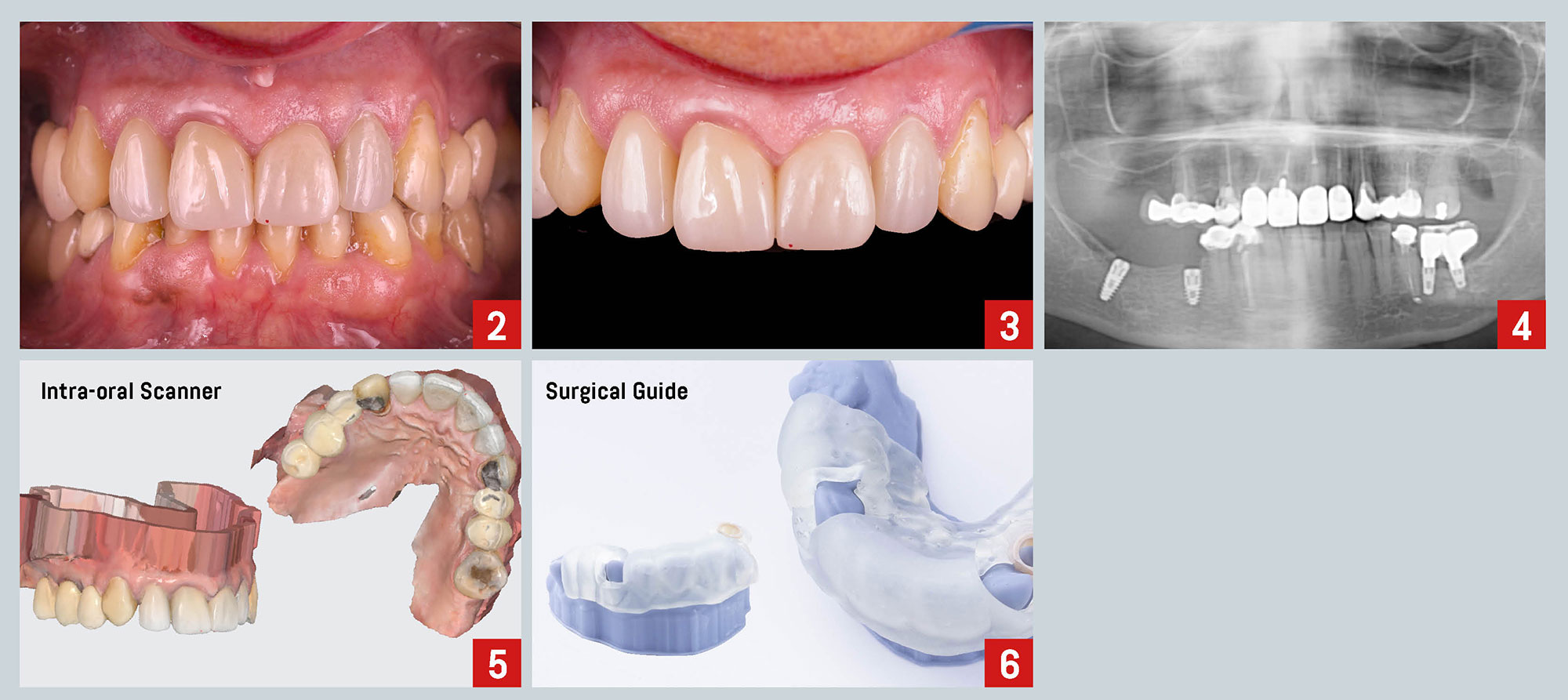
Following a proper diagnosis, the treatment plan proposed involved the extraction of hopeless teeth #1.3 and #1.5, and the placement of three implants, followed by the placement of immediate final abutments at the same time of the surgery (One Time Clever Concept, C-Tech Implant). The planned final prosthesis consisted of a single crown and a 3-unit bridge over implants.
 In 1997, it was published a study that demonstrated that repeated abutment disconnection/reconnection compromised the mucosal barrier resulting in the apical displacement of connective tissue attachment and additional marginal bone loss1. The One Time abutment concept aims to avoid disrupting the fragile soft tissue seal around implants and maximize marginal bone stability.
Initial photos of the patient’s current situation and a CBCT were taken. Also, initial upper and lower arch impressions were obtained using an intra-oral scanner (Fig. 2-5). The intra-oral scan and CBCT were superimposed using a specific 3D CAD software to plan implant placement. Once the implant positions were defined, they were translated into the design of the surgical guide (Fig. 6). The size of the one-time abutments was also planned in the same software as the implants.
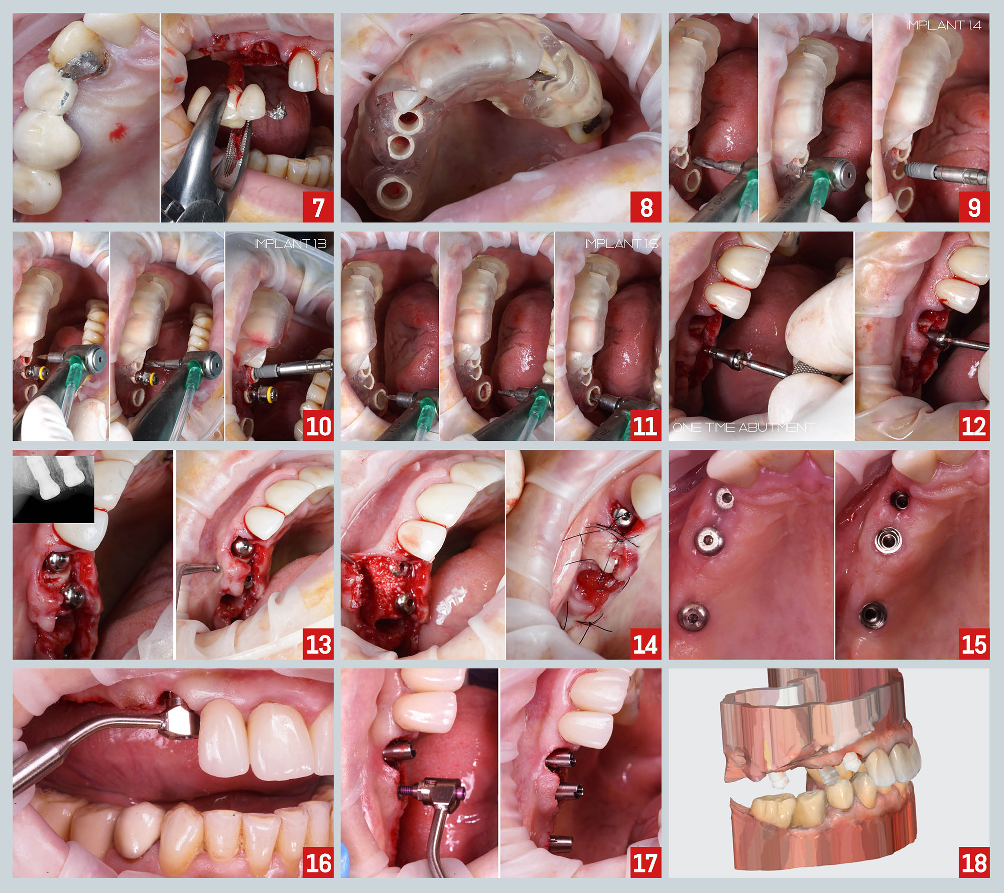
At the time of the surgery, extraction of teeth #1.3 and #1.5 were carried out, and the stability and adaptation of the guide were checked, followed by guided preparation of the implant sites according to a specific drilling protocol and using C-Tech guided surgery kit. Implants were placed fully guided on 1.3, 1.4, and 1.6 sites (Fig. 7-11). Then, One Time abutments with 3mm of gingival height were seated on the implants and fastened with the cover screw which works also as the healing abutment (Fig. 12-14).
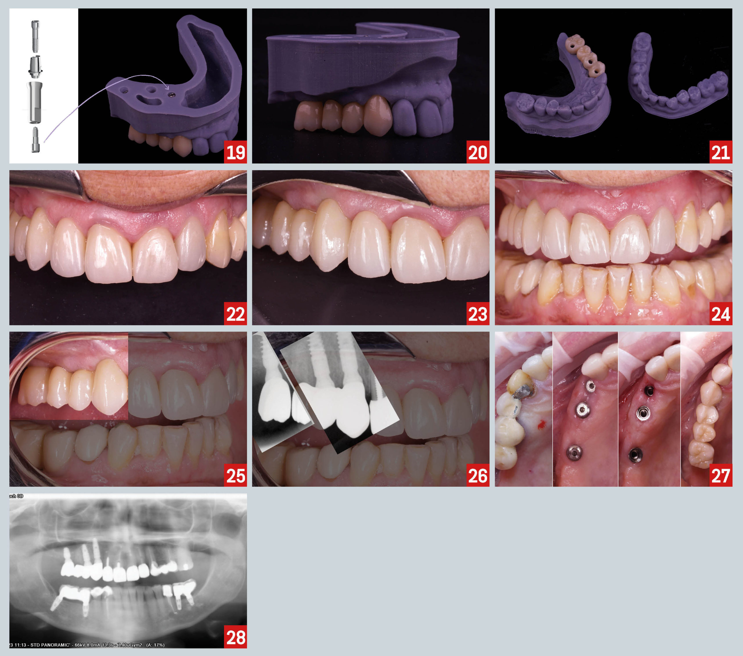 After the osseointegration period, final impressions were taken. First, the cover screw was removed and titanium bases were placed over the One Time abutments. Then, a digital impression was performed using scan caps (Fig. 15-18). The laboratory technician used CAD-CAM technology to obtain a 3D model and to design and mill the final restorations (Fig. 19-21). Finally, the 3-unit bridge and the single crown over implant were delivered and final records were obtained: intra-oral pictures, periapical and panoramic x-ray (Fig. 22-28).
1-Abrahamsson, I., Berglundh, T., & Lindhe, J. (1997). The mucosal barrier following abutment dis/reconnection. An experimental study in dogs. Journal of clinical periodontology, 24(8), 568- 572.