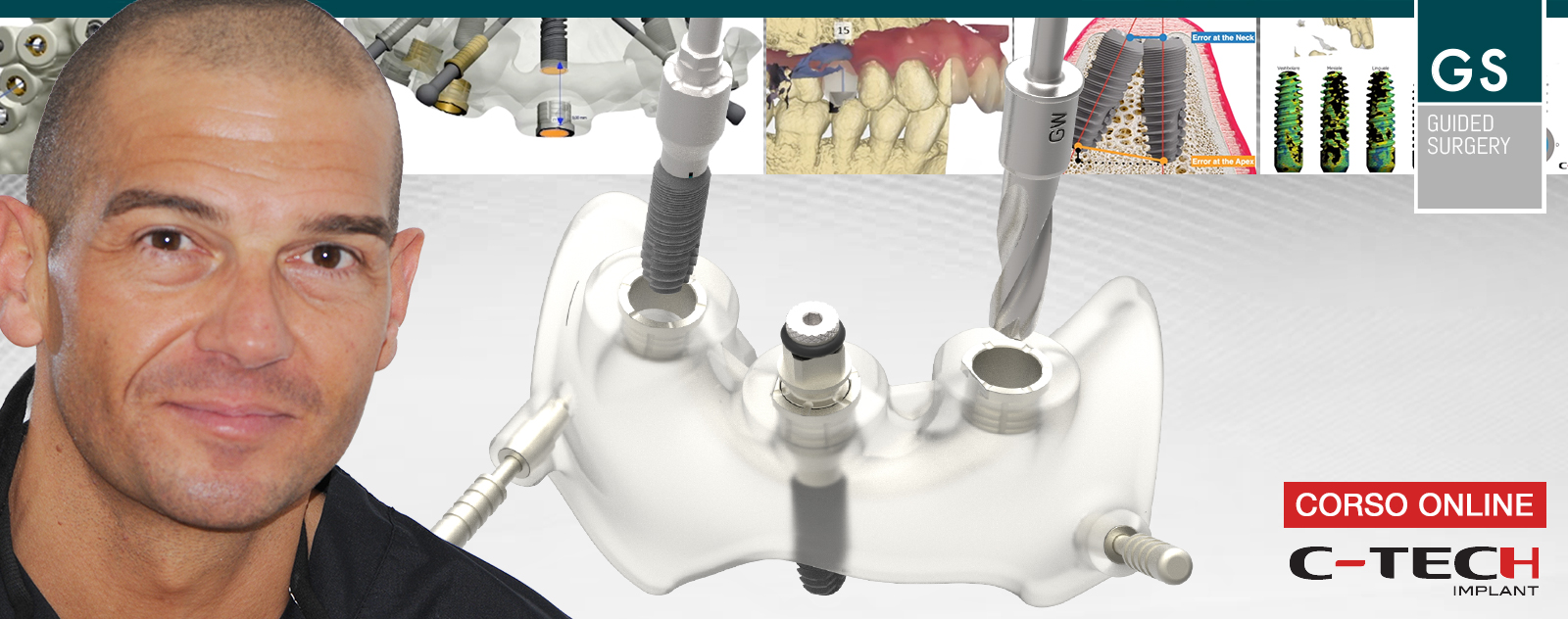
Guided Surgery Webinar | Digital implant planning: workflow and suggestions for the best results in guided surgery

by Dr. Luigi Ciacci, Dentist, expert in guided surgery, ODT Andrea Sessa

Dr Fabrizia Luongo, DDS, MS, Periodontist, Rome, Italy
Introduction
The evolution of digital dentistry and the development of a digital workflow has concentrated on digital planning with the use of Cone Beam CT scanning as well as using digital restorative tools to combine DICOM (Digital Imaging Communication in Medicine) and .stl (stereolithography) files to virtually plan, place and restore implants before using this plan to treat patients. The resulting benefits are reduced chair time, high precision and predictable aesthetic results often with immediate fixed provisional restorations available at time of surgery and corresponding high levels of patient satisfaction.
Intra-oral scanning to create digital ‘virtual impressions’ is also becoming more prevalent with the information being stored in the .stl file format. This information can be utilised by appropriate CAD/CAM (computer-aided design and computer aided manufacturing) software to design and manufacture a dental restoration (either by milling or 3D printing).
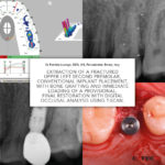
One area that is sometimes overlooked is the use of digital technology in occlusal analysis and adjustment of the restored dental implant. The following case study examines the occlusal management of a conventionally placed implant.
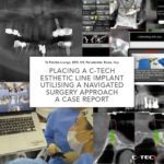
Dr Fabrizia Luongo, DDS, MS, Periodontist, Rome, Italy
Introduction
The use of Cone Beam Computerised Tomography (CBCT) scanning is becoming more commonplace in today’s implant dentistry. These scans combined with increasingly sophisticated software technology has led to greater accuracy in digital planning and guided surgery and is gradually being adopted across general practice. Furthermore, the restorative planning can be digitised and combined with the CBCT data in appropriate planning software.
In conjunction with these technology advances, dental implant design has evolved to incorporate the latest concepts of biomechanical design, which include sophisticated thread profiling, platform switching and a Morse locking taper on the implant/abutment interface.
The following case details brings these advances in implants and digital technologies together.
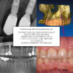
Dr Fabrizia Luongo, DDS, MS, Periodontist, Rome, Italy
Introduction
There has been a considerable evolution in implant dentistry over recent years that has seen the design of dental implants adopt sophisticated thread profiles which leads to a better primary stability. Simultaneously these new geometries aid cortical bone maintenance through to platform-switching capabilities again designed to minimise bone loss. When combined with improved implant-prosthetic connections such as a Morse-locking conical connection it contributes to a good long-term prognosis and an aesthetic outcome.
Concurrent with this evolution in implant design have been substantial advances in digital technologies across the field of dentistry. These digital advances include Cone-beam CT scanning combined with appropriate 3D planning software, navigated surgery technology, 3D intra-oral scanning to create a ‘virtual impression’ and 3D printing technologies. These digital technologies can minimise the number of appointments found in a conventional treatment protocol as well as enabling greater accuracy and will be considered in this case study.

dr Ivona Bjenjaš, Dental office BGD Osmeh, Belgrade, Serbia
Abstract
We described a case report of a 66 years old patient with asymptomatic impacted maxillary canine free of any surrounding pathology. Extraction of the impacted tooth was avoided because of expected massive bone loss and possible complications. Two C-Tech implants were placed through the impacted tooth in region 13 and 14. Primary stability was achieved on 60N/cm. No postoperative pain, swelling or bleeding was reported by the patient. After four months implants were uncovered and rehabilitated prosthetically with E-max CAD bridge.
We present you a Surgical Clinical Case with Flapless Technique performed by Doctor Aldo De Blasi using SD Mini Implants. You will be able to observe all the steps necessary for the stabilization of the lower prosthesis in the interforaminal region.

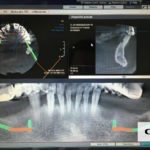
Clinical case of guided surgery by Doctor Marco Cernicchi. From computer design and model creation to the operation on the patient.
A 55 year old female patient was referred to our office for a full mouth rehabilitation. Upon clinical examination and a CBCT, we have decided to implant 4 EL C-Tech conical Morse tapered connection implants in the upper jaw for a bar retained over denture and due to financial considerations; was to perform extraction of all remaining lower teeth and immediate post extraction implantation of SD C-Tech mini dental implants. A full muco-periosteal flap was released, teeth were extracted, an alveotomy was performed to achieve an optimal bone platform for the SD mini dental implants and to obtain a bigger vertical dimension for the overdenture!
A 58 years old female patient has come to our clinic for lower jaw rehabilitation. After the clinical examination and OPG, We have decided to extract tooth 33, which is the only one remaining. As thepatient has not accepted a big augmentation procedure, we have decided to implant 4 C-Tech SD mini dental implants for retention and stabilization of the lower denture.