• Italiano
    • English
    • Deutsch
    • Español
    • Français
    • Русский
    • 中文 (中国)
    • Türkçe
    • Ελληνικά
    • Magyar
    • Српски језик
    • Lietuviškai
    • Bahasa Indonesia
    • Slovenčina

Slide Casi clinici

Регенерација Костију

Bridge on upper jaw with bone graft, 5-year follow-up

Written by feRKzzCTo0 on . Posted in ND – Narrow Diameter, Peng Dong, Клинички случајеви, Конвенционална Имплантологија, Регенерација Костију

Doctor Peng Dong, the expert of tooth implantation in Peking University International Hospital and president of Beijing Hedu Stomatological Clinic Co., Ltd

Medical Case Report

Sex / Age: Male/67  
First Visit: 16/05/2015 Implant Surgery: 03/07/2015
Others Surgery (GBR…):03/07/2015 Final Restoration: 26/12/2015
First Recall: 06/01/2016  
Chief Complaint: Loose fixed bridge on upper anterior teeth.
Consideration(s): GBR

CASE OUTLINE

Loose fixed bridge on upper anterior teeth. Carefully extract tooth 2, on the upper right side and tooth 1 on the upper left side. After the healing period, C-Tech implants will be placed in the sites of right upper tooth 2 and left upper tooth 1. After five months, an oxidized zirconia porcelain bridge will be used for the final restoration.

TREATMENT PLAN

For right upper tooth 2, the width of the alveolar bone is 7.6mm, and the height is 13.4mm. For left upper tooth 1, the width of the alveolar bone is 7.4mm, and the height is 14.5mm. C-Tech implants (Model: EL-4311) will be placed in both sites using conventional implantation with simple bone grafting. After a period of five months, the second phase of the surgery will be conducted.

TREATMENT

1. Extract venous blood for the preparation of CGF for later use.
2. Perform standard disinfection and drape the area with cloths. Administer painless local anesthesia to the upper front teeth region. Make horizontal incisions along the alveolar ridge from right upper tooth 2 to left upper tooth 1. Make incisions inside the grooves between right upper tooth 3 and left upper tooth 2. Perform a labial side vertical reduction on right upper tooth 3. Reflect the gingiva and thoroughly clean the wound, observing bone defects on the labial side of right upper tooth 2 and left upper tooth 1.
3. Precisely position the C-Tech implants (Model: EL-4311) in the locations of right upper tooth 2 and left upper tooth 1. Use tapered drills to create boreholes measuring 3.7*13mm for each implant. Implant the C-Tech implants with closure screws. Fill the bone defects and perforations on the labial side with a mixture of bone powder and CGF. Cover the gaps with membrane, and cover the alveolar ridge with another layer of membrane, followed by a layer of CGF membrane. Securely suture the area.
4. After six months, take an impression and replace the restorative abutments with Model: EL-4503F*2. Permanently fix them with adhesive fixation, and apply an oxidized zirconia porcelain bridge for long-lasting restoration.


Guided Implant Placement with Buccal Exposure and Graft – clinical case

Written by feRKzzCTo0 on . Posted in Alexandre Minis, CENTURY-GTR, GS – Guided Surgery, Вођена Хирургија, Клинички случајеви, Регенерација Костију

Dott. Alexandre Minis, France

Case description:
The 55 years old female patient has a lack of prior medical history and is a non-smoker.
There is a fracture under the crown of #12 with infection and vestibular fistula for which a temporary extraction with a valplast for 3 months is performed.
On the control CBCT, low bone volume and evidence of a radiolucent vestibular image initially suggesting fenestration linked to the fistula present before the extraction, or, of the bone being formed.

Surgical dental history:
The surgery is performed using a surgical guide, designed on 3Shape Implant Studio, dentition supported guide 3D printed on a Phrozen Shuffle printer.

Dentist Michael Weiland: excellent care is important to me!

Written by feRKzzCTo0 on . Posted in Scientific Publications, Вођена Хирургија, Конвенционална Имплантологија, Мини Импланти, Регенерација Костију

Magazine PIP • Practical implantology and implant prosthetics • 08 June 2022

Dentist Michael Weiland: excellent care is important to me!

In times of skilled labor bottlenecks and a fundamental lack of staff, the first thing that many lose is the services that were once seen as so essential. But the more a piece of hardware is interchangeable and discount battles want to be avoided, the more a company stands out from the customer with the many free little help.
Interview with dentist Michael Weiland and his team


pip: How did your first contact with the implant manufacturer C-Tech and Mr. Lütfü Agic come about?
Michael Weiland: I’ve known Mr. Agic for a very long time, for more than ten years, from my time in practice. Four years ago I set up my own practice, but at first I had too much to do with getting the practice up and running. At the same time, a lot was happening within our family – as it is at our age, that often happens alltogether. That’s why I didn’t deal with implantology myself at first and only provided prosthetics at first. Nevertheless, contact with Mr. Agic was never lost, especially since my basic interest continued to exist. Mr. Agic then drew my attention to the further training here at the municipal clinic with Prof. Anton Dunsche.

pip: What is particularly important to you at a training event?
Michael Weiland: Everyone is always talking about ‘take home messages’, and that’s actually what it’s all about. I would like to learn something that I can use in my practice in the relatively short term. In addition to the theory, which you can read up on or acquire through an online event, it is important to me to have the opportunity to try out and practice techniques yourself, preferably with someone at your side who can immediately give you one or two good ones can show tricks and tips. It’s about getting more security for me in my practice. The first course with Prof. Dunsche was just super organized, we learned theory, but also a lot of practice with exercises on the pig’s jaw and later the opportunity to assist Prof. Dunsche himself with surgeries.

pip: So it didn’t stop at this event?
Michael Weiland: No, after that I was given the opportunity to attend a four-day course in Palermo, Italy. They do things slightly differently there, with personal warmth and beautiful surroundings. Everything was very well organized, we even got two translators at our side, so there were no problems here either. For two days we were introduced to the C-Tech system and various surgical techniques, and we were able to use the third and fourth day very intensively for practical exercises and treatments directly on the patient. The two very experienced speakers also supervised this part and took over more complex situations myself, which I might never want to do myself in my practice, but which were exciting to watch. But I also learned an enormous number of new techniques for my areas of application and was able to practice them in such a way that I now feel safe with them. After all, I want to offer my patients the ‘state of the art’ of today’s implantology.


pip:
How important is the support provided by Mr. Agic to you?
Michael Weiland: We work directly with and on people, and I personally prefer it if I too am seen as a person. Mr. Agic has never promised too much, is always available and implements my requests incredibly quickly, even if I reach him on the way. I receive all important information promptly and in Mr. Agic I have a good partner for the exchange of ideas, at the same time I never feel pressured. I sometimes think that my time is more important to Mr. Agic than his own, and I can feel that through this great service.

Small but powerful. High-quality range with C-Tech system implants.

Written by feRKzzCTo0 on . Posted in El – Esthetic Line, Nd – Narrow Diameter, Scientific Publications, Вођена Хирургија, Конвенционална Имплантологија, Мини Импланти, Регенерација Костију

Magazine PIP • Practical implantology and implant prosthetics • November 2019

Small but powerful. High-quality range with C-Tech system implants.

Full Smile, under the direction of business owner Lütfü Agic, well-known in the field of dental implantology, advises on a high-quality range with C-Tech system implants, biomaterials such as Bioteck, as well as dental and surgical instruments and tools from devemed. How can such a small unit survive in times of ever greater global players in the field of dentistry and what is the reason for a dentist to look for the partnership, asked pip and talked to Stefan Grümer, M.Sc., who runs together with colleagues a large referral practice on Theaterstrasse in Aachen.

pip: Don’t you kind of lose touch with international developments and trends with such a small partner like Full Smile?
Dr. Grümer: That may well be, regarding these international trends. Here, in the Theaterstraße in Aachen, we are a practice with an impressive range of modern services, especially in the field of restorative and aesthetic dentistry. From 3D diagnostics and computer-based functional diagnostics to laser dentistry, we use state-of-the-art and exceptionally gentle treatment methods that are comfortable for the patient. We are focusing strongly on training programs so that our range of services stays up to date with the latest state of science and research. But we do not have to follow any pseudo innovation. Much more important to us is a certain consistency with our commercial partners, because as well as between us and our patients, the cooperation between us and a medical device manufacturer is a matter of trust. It would bother me to have to adjust to a new contact person every few months, who knows neither me nor my treatment philosophy nor knows what my experiences are and what might actually enrich and interest me. Since 2013 I have been running the first university teaching practice in Germany in cooperation with the RWTH Aachen, an international academy with many foreign and extensive research projects – above all in the field of implantology and laser dentistry. Therefore, we are demanding a certain level of consulting expertise and a portfolio at eye level from our partners.

pip: What impressed you most about the C-Tech Implant System?
Dr. Grümer: As a referral practice we naturally work with different systems. Personally, I especially appreciate the C-Tech Implant System because of its system-related features. It is a fully engineered system that provides different implant diameters and lengths for all common indications – up to special designs such as one-piece mini-implants for narrow spaces, abutments or anchoring of dentures. At C-Tech we experience a very high primary stability and a very easy handling and thus a method which is absolutely suitable for everyday use. Furthermore, the aesthetic demands of our patients have increased significantly. With the EL – Esthetic Line – C-Tech offers a special design for these more demanding challenges. Refinements, like the platform switch, a generous apical threading, the morse taper connection and the concave aesthetic concept create a good stability with excellent shaping of the surrounding tissue. Despite a high prosthetic design freedom, the system is very easy to use, but the prosthetic connection is identical to the other implant diameters of the series. So you can get by with a straightforward instrumentation. I also like a whole new service of Full Smile, which I have never experienced before: As a dentist, I can search for any product via Full Smile and, if I found one, I will only pay the same list price as in the catalogue!

pip: What about service and accessibility – how can such a small company compete with companies that are quite different staffed?
Dr. Grümer: In fact, we sometimes ask ourselves the famous question of how often Mr. Agic has been cloned. He’s really frequently on the move and also visits us regularly to exchange ideas with us or to prepare ourselves for a new technique. However, we always reach someone in the office or receive a callback within a very short time – and also in such a short time our request is accepted, a delivery is initiated or a technical question is answered. In fact, with other companies I sometimes spent a lot more time in some telephone loop with automated speech before I can speak to a human being. With individual consultation, the reliable assurance of the desired delivery times and practical training, Full Smile is a professional partner for us in the field of dental implantology, periodontology and surgery.


Italian passion for German precision

Written by feRKzzCTo0 on . Posted in El – Esthetic Line, Nd – Narrow Diameter, Scientific Publications, Sd-Mb – Monoblock - Small Diameter, Вођена Хирургија, Конвенционална Имплантологија, Мини Импланти, Регенерација Костију

Magazine PIP • Practical implantology and implant prosthetics • August 2018

Italian passion for German precision

When visiting the impressive manufacturing facility, with 25 CNC machines alone, you will also come across the “Mercedes” of manufacturing technology such as the Swiss Tornos, a five-axis machine, which at C-Tech is purely tasked with the manufacture of mini-implants. Currently they are the largest manufacturer in Europe for this specific type of implant. Mini-implants are popular as a minimally-invasive option for creating an immediate restoration, which is currently a highly sought-after alternative for fast and good functional care of older patients. An impressive neighbour is the Willemin Macodell (the Rolls-Royce of CNC machines), which can be fitted with 48 different CNC cutters. It can change tools in 1.4 seconds and can be used in any conceivable axis, including for the manufacture of angled mini-abutments. C-Tech was one of the first companies to move over to cooling all computer-controlled machine tools using water-miscible bio-lubricants. The plant-based oil is odour-free, improves machining and even has a wider range of applications than mineral oil products. “And even though of course we run extensive cleaning processes for the finished parts, it’s still one risk factor less”, comments Lütfü Agic with respect to the recently published independent surface study by the University of Cologne, where the C-Tech implant surfaces were tested as an example, in testing applied to very few dental implant surfaces on the dental market.

A one year follow up examining bone level preservation utilising platform switching implants – Ningxia Med J Dec. 2017

Written by feRKzzCTo0 on . Posted in Century-Gtr, Scientific Publications, Конвенционална Имплантологија, Регенерација Костију

Ningxia Med J, Dec. 2017, Vol 39, No. 12

A one year follow up examining bone level preservation utilising platform switching implants

Jianlin Chen, Yuanjie Cao, Lili Shan, Yan Li, Li Ma, Na Wang, Xiuyu Wu Author’s
Unit: Department of Stomatology, Lingwu People’s Hospital, Ningxia

Abstract

Objective The aim of this research is to observe and evaluate the clinical application of the Esthetic Line [EL] implant system (C-Tech, Bologna, Italy). The EL implant has a surface topography created by sandblasting and acid etching (SLA) and a Morse locking conical connection.

Methods 35 patients with one or more missing teeth were selected and a total of 60 EL implants were placed using either a one stage (non-submerged) placement protocol or a two stage (submerged) protocol. Where applicable, second stage surgery was undertaken 2 ~ 4 months post implantation. Subsequent to this the EL implants were permanently restored over a period of 2 ~ 4 weeks. Clinical examination and imaging analysis were undertaken to evaluate clinical success.

Results All 60 implants osseointegrated successfully, and at the one year follow up there was a 100% retention rate and no adverse reactions in the host. Mesial and distal bone heights were recorded on the day of surgery as well as at the fitting of the permanent restoration and after the implants had been functionally loaded for 12 months. Mesial bone heights were (0. 35 ± 0. 49) mm, (0. 18 ± 0. 44) mm and (0. 25 ± 0. 36) mm respectively. Distal bone heights were (0. 20 ± 0. 42) mm, (0. 08 ± 0. 45) mm and (0. 15 ± 0. 38) mm. In the first year of implant functional load, the total absorption of the mesial bone was (- 0. 11 ± 0. 38) mm and the distance was (- 0. 07 ± 0. 31) mm; There was no significant difference in blood indexes between the preoperative and postoperative three months (P < 0. 05).

Conclusion The design of the EL implant incorporating a combination of a parallel walled section with an apical taper, a beveled shoulder, platform switching, a Morse locking conical connection combined with an SLA treated surface and a sophisticated double threaded morphological design all contribute to successful treatment.

Key words Italy; C – Tech Esthetic Line [EL] implant; implant design; surface treatment; Morse-locking.

In recent years, the evolution of implant techniques combined with developments in implant design and restorative techniques has resulted in greater acceptance of implant treatment by the majority of patients. The Stomatology department of our hospital has adopted the Esthetic Line [EL] implant system (C-Tech, Bologna, Italy) for use in our clinic. A study into the clinical results of 60 EL implants placed in 35 patients was undertaken.  

Conclusions

To sum up, the innovative design of the Esthetic Line implant with its sequential thread design, micro-threads to preserve bone at the collar, sophisticated self-cutting and double lead threading preserves bone structure and increases bone to implant contact. This is combined with a SLA surface treatment, a combination of a bevelled shoulder with a platform switching design and a Morse locking conical connection to make it safe and effective in clinical application. The limitation of this study is that the number of cases included is relatively small, only 30 implants of 35 patients were followed up for 12 months. More accurate clinical effect requires more long-term retrospective and prospective clinical observation and research on larger sample size.

References

[1] Albrektsson T,Zarb G,Worthington P,et al. The long – termefficacy of currently used dental implants: a review and proposed criteria of success[J]. The International Journal of Oral & Maxillofacial Implants,1986,1 (1):11-25.

[2] 罗佳,胡秀莲,林野,等. 下后牙区平台转移设计种植体植入深 度对边缘骨水平影响的临床研究[J]. 北京大学学报(医学 版),2012,44 (1):65-69.

[3] 张剑明,郭平川,刘春年,等. 口腔种植修复技术在无牙颌患者 中的应用[J]. 中国口腔种植学杂志,2009,14( 2) : 64 – 65. 1. 陶江丰,陈宁,周芷萱. 口腔种植领域中再生医学的研究进展 [J]. 口腔医学,2012,32(7):443-446.

[4] Kim YK,Kim YJ,Yun PY,et al. Effects of the taper shape,dual- thread and length on the mechanical properties of mini – implants [J]. Angle Orthodontist, 2009, 79(5):908 – 914. 

[5] Hnang HL,Chang CH,Hsu JT,et al. Comparison of implant body designs and threaded designs of dental implants: a 3 – dimensional finite element analysis[J]. In J Oral Maxillofac Implants,2007,22 (4):551-562. 

[6] Bolind K,Johansson CB,Becker W,et al. A descriptive study on re- trieved non – threaded and threaded implant designs[J]. Clinical Oral Implants Research, 2005, 16(4):447 – 455. 

[7] 付丽,周延民. 平台转换结构中肩台变化对种植体 – 骨界面应 力分布的影响[J]. 中国口腔种植学杂志, 2009, 14(2):120.

[8] 郭智舜,郭平川,周贵祥,等. 平台转换种植体肩台宽度对周围 组织的影响[J]. 实用口腔医学杂志, 2013, 29(3):352 – 357.

[9] 宿玉成. 口腔种植学[M]. 第 2 版. 北京: 人民卫生出版社, 2014: 349.

[10] 丁旭,汪大林. 影响种植体及其周围组织应力分布的原因分析 [J]. 口腔颌面修复学杂志, 2008, 9(2):155 – 157.

[11] 董福生, 董玉英,邢汝东,等. 种植体长度对骨界面应力分布影 响 的 三 维 有 限 元 分 析 [ J ] . 中 国 口 腔 种 植 学 杂 志, 2001, 6 (3):106-108.

[12] Nikellis I, Levi A, Nicolopo UC. Immediate loading of 190 endos- seous dental implants: a prospective observational study of 40 pa- tient treatments with up to 2 – year data[J]. The International Journal of Oral & Maxillofacial Implants, 2004, 19(1):116-123.

[13] 宿玉成. 现代口腔种植学[M]. 北京: 人民卫生出版社, 2004: 63-71.

[14] Elias CN, Meirelles L. Improving osseoin tegration of dental implant [J]. Expert Review of Medical Devices,2010,7(2):241-256.

[15] Le Guehennec L,Soueidan A,Layrolle P,et al. Surface treatments of Titanium dental implants for rapid osseointegration[J]. Dental Materials,2007,23(7):844-854.

[16] 刘长虹,杨晓喻. 钛种植体表面特征对种植体周细胞功能的影 响[J]. 中国口腔种植学杂志,2008, 13(2):81-84.

[17] Saivi GG,Gallini G,Lang NP. Early loading (2 or 6 weeks) of sandblasted and acid – etched( SLA) ITI impland in the posterior mandible[J]. Clin Oral Implres,2004,15(1):142-149.

[18] 郭苏伟,赵保东,刘凤芝,等. 平台转换技术对上颌前牙区单枚 种植修复影响的临床观察[J]. 中国口腔种植学杂志,2011,16 (2):120-124.

[19] Otto M. Concept of implant switching [J]. Journal of the South African Dental Association, 2007, 62(2):80.

[20] Lazzara RJ, Porter SS. Platform switching: a new concept in implant dentistry for controlling postrestorative crestal bone levels[J]. The International Journal of Periodontics & Restorative Dentistry, 2006, 26 (1):9 – 1 7 .

[21] 林野. 当代口腔种植学的进展及其临床意义[J]. 口腔颌面外 科杂志, 2006, 16(4):285-290.

[22] 张志勇,王慧明,赖红昌. 口腔颌面种植修复学[M]. 上海: 世 界图书出版公司, 2009: 54.

International Journal of Implant Dentistry – August 2017 – Investigation of peri-implant tissue conditions and peri-implant tissue stability in implants placed with simultaneous augmentation procedure: a 3-year retrospective follow-up analysis of a newly developed bone level implant system

Written by feRKzzCTo0 on . Posted in Century-Gtr, El – Esthetic Line, Henriette Lerner, Robert Sader, Scientific Publications, Shahram Ghanaati, Конвенционална Имплантологија, Регенерација Костију

International Journal of Implant Dentistry • August 2017

Investigation of peri-implant tissue conditions and peri-implant tissue stability in implants placed with simultaneous augmentation procedure: a 3-year retrospective follow-up analysis of a newly developed bone level implant system

Jonas Lorenz University Hospital Frankfurt · Department of oral, maxillofacial and plastic surgery, Henriette Lerner HL DENTCLINIC, Robert A. Sader Goethe-Universität Frankfurt am Main · Center of Stomatology
and Shahram Ghanaati Goethe University of Frankfurt/Main; Universitätsmedizin der Johannes Gutenberg-Universität Mainz · Department for Oral, Craniomaxillofacial and Facial Plastic Surgery; Institute of Pathology

The aim of the present retrospective analysis was to assess peri-implant tissue conditions and document peri-implant tissue stability in C-Tech implants when placed simultaneously with a GBR (Guided Bone Regeneration) augmentation procedure.

A total of 47 implants, which were placed simultaneously with a GBR (Guided Bone Regeneration) procedure with a synthetic bone substitute material in 20 patients, were investigated clinically and radiologically at least 3 years after loading.

The follow-up investigation revealed a survival rate of 100% and only low median rates for probing depths (2.7 mm) and BOP (bleeding on probing) (30%). The mean PES (Pink Esthetic Score) was 10.1 from the maximum value of 14.
No osseous peri-implant defects were obvious, and the mean bone loss was 0.55 mm.

In conclusion, implants placed in combination with a GBR (Guided Bone Regeneration) procedure can achieve long-term stable functionally and esthetically satisfying results for replacing missing teeth in cases of atrophy of the alveolar crest.

References

1. Gurgel BC, Montenegro SC, Dantas PM, Pascoal AL, Lima KC, Calderon PD. Frequency of peri-implant diseases and associated factors. Clin Oral Implants Res. 2016; doi: 10.1111/clr.12944

2. Qian J, Wennerberg A, Albrektsson T. Reasons for marginal bone loss around oral implants. Clin Implant Dent Relat Res. 2012;14(6):792–807.

3. Berglundh T, Lindhe J, Ericsson I, Marinello C, Liljenberg B, Thomsen P. The soft tissue barrier at implants and teeth. Clin Oral Implants Res. 1991;2:81–90.

4. Berglundh T, Lindhe J, Jonsson K, Ericsson I. The topography of the vascular systems in the per iodontal and peri-implant tissues in the dog. J Clin Periodontol. 1999;21:189–93.

5. Moon I, Berglundh T, Abrahamsson I, Linder E, Lindhe J. The barrier between the keratinized mucosa and the dental implant. An experimental study in the dog. J Clin Periodontol. 1999;26:658–63.

6. Lindhe J, Berglundh T. The interface between the mucosa and the implant. Periodontol. 1998;17:47–54.

7. Masaki C, Nakamoto T, Mukaibo T, Kondo Y, Hosokawa R. Strategies for alveolar ridge reconstruction and preservation for implant therapy. J Prosthodont Res. 2015;59(4):220–8.

8. Damien CJ, Parsons JR. Bone graft and bone graft substitutes: areview of current technology and applications. J Appl Biomater.1991;2:187–208.

9. Cordaro L, Torsello F, Miuccio MT, di Torresanto VM, Eliopoulos D. Mandibular bone harvesting for alveolar reconstruction and implant placement: subjective and objective cross-sectional evaluation of donor and recipient site up to 4 y
ears. Clinical Oral Impl Res. 2011;22:1320–6.

10. Canullo L, Penarrocha-Oltra D, Soldini C, Mazzocco F, Penarrocha M, Covani U. Microbiological assessment of the implant-abutment interface in different connections: cross-sectional study after 5 years of functional loading. Clin Oral Implants Res. 2015;26(4):426–34.

11. Misch C. Implant design considerations for the posterior regions of the mouth. Implant Dent. 1999;8(4).

12. SteigengaJ,al-ShammariK,NocitiF,MischC,WangH.Dentalimplant design and its relationship to long-term implant success. Implant Dent. 2001;12(4):306–17.

13. Canullo L, Pace F, Coelho P, Sciubba E, Vozza I. The influence of platform switching on the biomechanical aspects of the implant-abutment system. A three dimensional finite element study. Med Oral Patol Oral Cir Bucal. 2011;16(6):852–6.

14. Lerner H, Lorenz J, Sader R, Ghanaati S. Two-year retrospective study of periimplant health and periimplant bone stability after immediate implant placement of a newly developed bone level implant system—a first report.
EDI Journal (European Association of Dental Implantologists, Teamwork Media); 2017; ahead of print.

15. Ghanaati S, Lorenz J, Obreja K, Choukroun J, Landes C, Sader R. Nanocrystalline hydroxyapatite-base d material already contributes to implant stability after 3 months: a clinical and radiologic 3-year follow-up investigation. In: Journal of Or al Implantology. 2014;40(1):103–9.

16. Lorenz J, Kubesch A, Korzinskas T, Barbeck M, Landes C, Sader R, et al. TRAP-positive multinucleated giant cells are foreign body giant cells rather than osteoclasts: results from a split-mouth study in humans. J Oral Implantol. 2015;41(6):e257–66.

17. Barbeck M, Udeabor S, Lorenz J, Schlee M, Grosse Holthaus M, Raetscho N, et al. High-temperature sintering of xenogeneic bone substitutes leads to increased multinucleated giant cell formation: in vivo and preliminary clinical results. J Oral Implantol. 2015;41(5):e212–22.

18. Barbeck M, Udeabor S, Lorenz J, Kubesch A, Choukroun J, Sader R, et al. Induction of multinucleated giant cells in response to small sized bovine bone substitute (Bio-Oss TM) results in an enhanced early implantation bed vascularization. Ann Maxillofac Surg. 2014;4(2):150–7.

19. Lorenz J, Barbeck M, Sader R, Russe P, Choukroun J, Kirkpatrick CJ, et al. Foreign body giant cell related encapsulation of a synthetic material three years after augmentation. J Oral Implantol. 2016;42(3):273–7.

School of Dental Medicine University of Belgrade, Serbia – November 2015 – Clinical outcomes following horizontal ridge augmentation with bone block grafts

Written by feRKzzCTo0 on . Posted in Century-Gtr, Scientific Publications, Конвенционална Имплантологија, Регенерација Костију

School of Dental Medicine University of Belgrade, Serbia • November 2015

Clinical outcomes following horizontal ridge augmentation with bone block grafts

Assoc Prof Dr Snezana Colic (School of Dental Medicine University of Belgrade, Serbia)

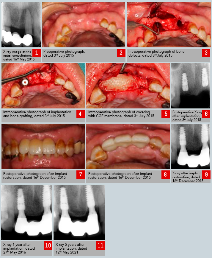
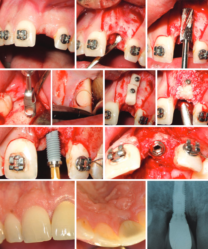
The aim of this study was to analyze clinical outcome of horizontal augmentation with autologous bone block grafts for the reconstruction of narrow edentulous ridge before implant placement. Eighteen partially edentulous patients, presenting insufficient bone width (less than 4mm) in the sites for implant placement were selected.
One or multiple cylindrical block grafts were harvested with trephine burrs from retromolar region and stabilized with titanium miniscrews. Fixed grafts were covered with deproteinised bovine bone mineral granules (DBBM) and collagen membrane (CM). The average amount of bone gain was 3,6 mm. One of the 24 block grafts was lost during the early healing period. Five months later, during the re-entry for implants placement, the gain of ridge width obtained were measured. All implants were placed in correct position and considered successfully integrated during the observation period. Three month after the implant placement prosthetic rehabilitation was started.
The mean follow up after prosthetic load has been 25,4 months.

This technique is reliable means for the correction of narrow edentulous ridge. Close contact between the bone graft and recipient bed surface ensure fast and intense vascularization and subsequent osseointegration of the graft. Adding bovine bone mineral and collagen membrane over bone block minimize resorption during healing.
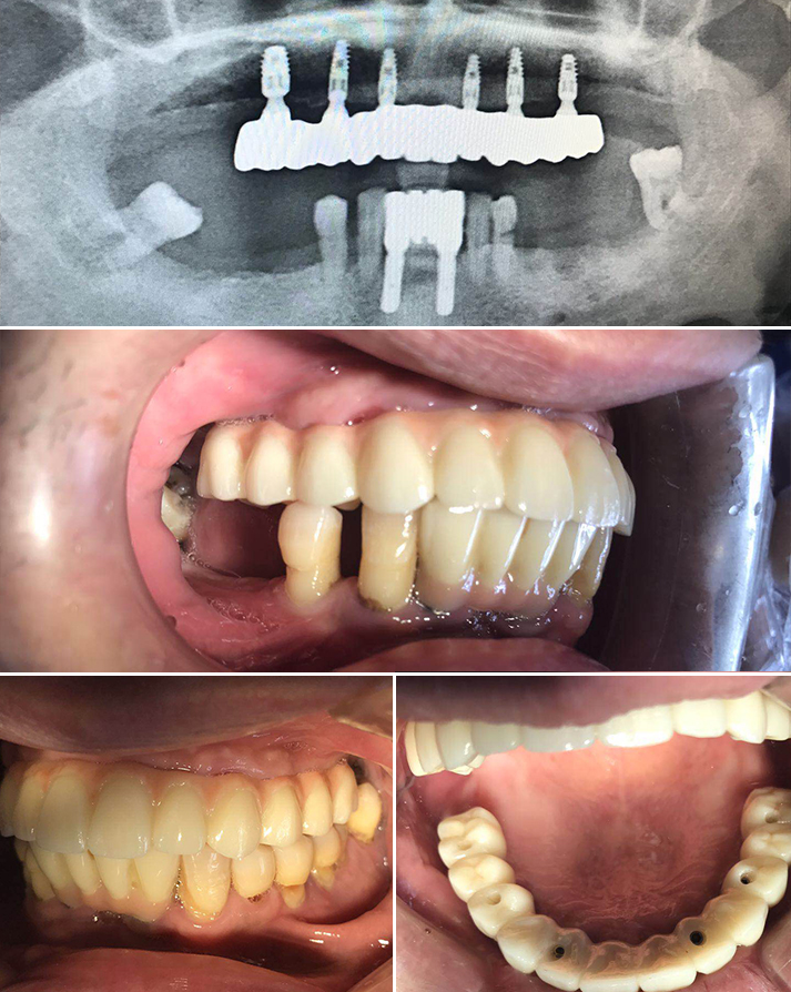
In all cases all planned implants could be placed in the correct positions.
The mean duration of follow up of the implants was 25,4 months. Prosthodontics rehabilitation involved single crowns or bridges.
Not a single implant was lost during the observation period. All implants were successful both aesthetically and functionally.


References

1. Albrektsson T., Zarb G., Wothington P. & Ericsson A.R. (1986) The long-term efficacy of currently used dental implants: A review and proposed criteria of success. International Journal of Oral and Maxillofacial Implants 1:1-25.

2. Araujo, M.G., Sonohara, M., Hayacibara, R., Car- daropoli, G.
& Lindhe, J. (2002) Lateral ridge augmentation by the use of grafts comprised of autologous bone or a biomaterial. An experiment in the dog. Journal of Clinical Periodontology 29:1122–1131.

3. Buser, D., Dula, K., Belser, U. C., Hirt, H. P. & Berthold, H.
(1995) Localized ridge augmen- tation using guided bone regeneration. II. Surgical procedure in the mandible. International Journal of Periodontics and Restora- tive Dentistry 15,10–29.

4. Buser D., Dula K., Hirt H.P., & Schenk R. (1996) Localized ridge augmentation using autografts and barrier membranes: A clinical study with 40 partially edentulous patients. Journal of Oral and Maxillofacial Surgery 54:420-432

5. Buser, D., Dula, K., Hess, D., Hirt, H.P. & Belser, U.C. (1999) Localized ridge augmentation with autografts and barrier membranes. Periodontology 2000 19:151–163.

6. Buser, D., Ingimarsson, S., Dula, K., Lussi, A., Hirt, H. P. & Belser, U. C. (2002) Long-term stability of osseointegrated implants in aug- mented bone: a 5-year prospective study in partially edentulous patients. International Journal of Periodontics and Restorative Dentistry 22,109–117.

7. Chiapasco M, Romeo E & Vogel G. (1998) Three-dimensional reconstruction of a knife-edge edentulous maxilla by sinus elevation, onlay grafts and sagittal osteotomy of the anterior maxilla: preliminary surgical and prosthetic results. Journal of Oral and Maxillofacial Implants, 13:394-399

8. Chiapasco M., Abati S., Romeo E., Vogel G. (1999) Clinical outcome of autogenous bone blocks or guided bone regeneration with e-PTFE membranes for the reconstruction of narrow edentulous ridges. Clin Oral Impl Res 1999:10:278-288

9. Chiapasco, M., Zaniboni, M. & Boisco, M. (2006) Augmentation procedures for the rehabilitation of deficient edentulous ridges with oral implants. Clin- ical Oral Implants Research 17 (Suppl.):136–159.

10. Cordaro, L., Amade, D. S. & Cordaro, M. (2002) Clinical results of alveolar ridge aug- mentation with mandibular block bone grafts in partially edentulous patients prior to implant placement. Clinical Oral Implants Research 13,103–111.

11. Cordaro, L., Torsello, F., Accorsi Ribeiro, C., Libera- tore, M. & Mirisola di Torresanto, V.M. (2010) Inlay– onlay grafting for three-dimensional reconstruction of the posterior atropic maxilla with mandibular bone. The International Journal of Oral and Maxillofacial Surgery 39:350–357.

12. Cordaro L., Torsello F., Morcavallo S, Mirisola di Torresanto V. (2011) Effect of bovine bone and collagen membranes on healing of mandibular bone blocks: a prospective randomized controlled study. Clin. Oral Impl. Res. 22,2011;1145–1150.

13. Ha ‘mmerle, C.H. & Karring, T. (1998) Guided bone regeneration at oral implant sites. Periodontology 2000 17:151–175.

14. Jensen SS, Aaboe M, Pinholt EM, Hjorting-Hansen E, Melsen F, Ruy- ter IE. Tissue reaction and material characteristics of four bone substitutes. Int J Oral Maxillofac Implants 1996:11:55– 66.

15. Kuboki, Y., Jin, Q., Kikuchi, M., Mamood, J. & Takita, H. (2002) Geometry of artificial ECM: sizes of pores controlling phenotype expression in BMP-induced osteogenesis and chondrogenesis. Connective Tissue Research 43:529–534.

16. Lekholm U & Zarb G.A. (1995) Patient selection and preparation. In: Tissue-integrated prostheses: Osseointegration in clinical dentistry. Pp. 199-209. Chicago: Quintessence

17. Machtei, E.E. (2001) The effect of membrane ex- posure on the outcome of regenerative procedures in humans: a meta-analysis. Journal of Perio- dontology 72:512–516.

18. Misch, C. M. (1997) Comparison of intraoral sites for onlay grafting prior to implant placement. Journal of Oral and
Maxillofacial Implants, 6:767-776

19. Maiorana, C., Beretta, M., Salina, S. & Santoro, F. (2005) Reduction of autogenous bone graft resorption by means of bio-oss coverage: a prospective study. International Journal of Periodontics and Restorative Dentistry 25:19–25.

20. McAllister, B.S. & Haghighat, K. (2007) Bone aug- mentation techniques. Journal of Periodontology 78:377–396.

21. Nkenke, E., Schultze-Mosgau, S., Radespiel-Tro ‘ger, M., Kloss, F. & Neukam, F.W. (2001) Morbidity of harvesting of chin grafts: a prospective study. Clinical Oral Implants Research 12:495–502.

22. Sander, L., Frandsen, E.V., Arnbjerg, D., Warrer, K. & Karring, T. (1994) Effect of local metronidazole application on periodontal healing following guided tissue regeneration. Clinical findings. Journal of Periodontology 65:914–920.

23. Schwarz F., Ferrari D., Balic E., Buser D., Becker J., Sager M. Lateral ridge augmentation using equine- and bovine-derived cancellous bone blocks: a feasibility study in dogs. Clin. Oral Impl. Res. 21,2010;904–912.

24. Simion M., Baldoni M., Rossi P. & Zaffe D. (1994) A comparative study of the effectiveness of a e-PTFE membranes with and without early exposure during healing period. International Journal of Periodontology and Restorative Dentistry 14:167-180

25. Von Arx, T & Buser, D. (2006). Horizontal ridge augmentation using autogenous block grafts and the guided bone regeneration technique with collagen membranes: a clinical study with 42 patients. Clinical Oral Implants Research 17:359–366

(BDIZ EDI) Implant-Study 2014/2015 – March 2015 – Quantitative and qualitative element-analysis of implant-surface by SEM and EDX

Written by feRKzzCTo0 on . Posted in Scientific Publications, Конвенционална Имплантологија, Регенерација Костију

(BDIZ EDI) Implant-Study 2014/2015 • March 2015

Quantitative and qualitative element-analysis of implant-surface by SEM and EDX

Prof. Dr. Dr. Joachim E. Zöller Interdisciplinary Polyclinic for Oral Surgery and Implantology Dept. for Cranio Maxillofacial and Plastic Surgery, Dr. med. dent. Dirk U. Duddeck Head of Surface Analyses, In cooperation with the European Association of Dental Implantologists BDIZ-EDI Quality & Research Committee

Background and Aim
Implant surfaces are modified by microstructures and surface extension to improve osseointegration. Numerous studies showed an increased adhesion and osteoblastic matrix-production on retentive titanium surfaces.

In 2008 the University of Cologne, Germany and the BDIZ EDI (European Association of Dental Implantology) with its Quality and Research (Q&R) Committee (www.bdizedi.org) performed a scanning electron microscopic study and analyzed the surfaces of 23 enossal titanium implants of several manufacturers at the Interdisciplinary Policlinic for Oral Surgery and Implantology, Department for Craniomaxillofacial and Plastic Surgery, University Cologne(1). The tested implants showed isolated and/or extensive deposits. Depending on manufacturing process, accumulations of organic material (carbon) or inorganic material like aluminum, silicon, phosphor, sulfur, chlorine, potassium and calcium were found.

In 2011-2012 we performed the same protocol on 57 dental implants from different manufacturers. However, the manufacturing of implants requires an adequate system of quality controls. Although some manufacturers have made substantial improvements since our first survey in 2008, the study in 2011-2012 again singled out a few implants with larger areas of surface blasting residue and selective organic impurities(2).

The BDIZ EDI(3), representing more than 5,500 active implantologists in Europe, was asked in its general meeting to continue these analyses periodically and to publish the results in the European EDI Journal.

While using the same material and methods regarding the technical setup this study allows comparisons to the results of previous studies.

As a continuance of the two studies cited above the aim of study is to verify improvements of manufacturing and quality management as well as to demonstrate the high quality level of the participating manufacturers and implant companies.

1)Duddeck DU; Comparative investigation of various implant surfaces by SEM-Analysis. (Posterpresentation) 18th Annual Scientific Meeting of the European Association of osseointegration, 30 September-3 October 2009, Monaco, France

2)Duddeck, DU. et. al; Surface characteristics and quality of implants in sterile packaging, EDI Journal 2013-1

3)The BDIZ EDI has set the preliminary standards for a qualifying procedure of all implant systems and maintains structured continuing education. The primary tasks of BDIZ EDI are to provide members with support and advice and to improve the quality of implant materials

Preefooter SER