• Italiano
    • English
    • Deutsch
    • Español
    • Français
    • Русский
    • 中文 (中国)
    • Türkçe
    • Ελληνικά
    • Magyar
    • Српски језик
    • Lietuviškai
    • Bahasa Indonesia
    • Slovenčina

Slide Casi clinici

CENTURY-GTR

Guided Implant Placement with Buccal Exposure and Graft – clinical case

Written by feRKzzCTo0 on . Posted in Alexandre Minis, CENTURY-GTR, GS – Guided Surgery, Вођена Хирургија, Клинички случајеви, Регенерација Костију

Dott. Alexandre Minis, France

Case description:
The 55 years old female patient has a lack of prior medical history and is a non-smoker.
There is a fracture under the crown of #12 with infection and vestibular fistula for which a temporary extraction with a valplast for 3 months is performed.
On the control CBCT, low bone volume and evidence of a radiolucent vestibular image initially suggesting fenestration linked to the fistula present before the extraction, or, of the bone being formed.

Surgical dental history:
The surgery is performed using a surgical guide, designed on 3Shape Implant Studio, dentition supported guide 3D printed on a Phrozen Shuffle printer.

A one year follow up examining bone level preservation utilising platform switching implants – Ningxia Med J Dec. 2017

Written by feRKzzCTo0 on . Posted in Century-Gtr, Scientific Publications, Конвенционална Имплантологија, Регенерација Костију

Ningxia Med J, Dec. 2017, Vol 39, No. 12

A one year follow up examining bone level preservation utilising platform switching implants

Jianlin Chen, Yuanjie Cao, Lili Shan, Yan Li, Li Ma, Na Wang, Xiuyu Wu Author’s
Unit: Department of Stomatology, Lingwu People’s Hospital, Ningxia

Abstract

Objective The aim of this research is to observe and evaluate the clinical application of the Esthetic Line [EL] implant system (C-Tech, Bologna, Italy). The EL implant has a surface topography created by sandblasting and acid etching (SLA) and a Morse locking conical connection.

Methods 35 patients with one or more missing teeth were selected and a total of 60 EL implants were placed using either a one stage (non-submerged) placement protocol or a two stage (submerged) protocol. Where applicable, second stage surgery was undertaken 2 ~ 4 months post implantation. Subsequent to this the EL implants were permanently restored over a period of 2 ~ 4 weeks. Clinical examination and imaging analysis were undertaken to evaluate clinical success.

Results All 60 implants osseointegrated successfully, and at the one year follow up there was a 100% retention rate and no adverse reactions in the host. Mesial and distal bone heights were recorded on the day of surgery as well as at the fitting of the permanent restoration and after the implants had been functionally loaded for 12 months. Mesial bone heights were (0. 35 ± 0. 49) mm, (0. 18 ± 0. 44) mm and (0. 25 ± 0. 36) mm respectively. Distal bone heights were (0. 20 ± 0. 42) mm, (0. 08 ± 0. 45) mm and (0. 15 ± 0. 38) mm. In the first year of implant functional load, the total absorption of the mesial bone was (- 0. 11 ± 0. 38) mm and the distance was (- 0. 07 ± 0. 31) mm; There was no significant difference in blood indexes between the preoperative and postoperative three months (P < 0. 05).

Conclusion The design of the EL implant incorporating a combination of a parallel walled section with an apical taper, a beveled shoulder, platform switching, a Morse locking conical connection combined with an SLA treated surface and a sophisticated double threaded morphological design all contribute to successful treatment.

Key words Italy; C – Tech Esthetic Line [EL] implant; implant design; surface treatment; Morse-locking.

In recent years, the evolution of implant techniques combined with developments in implant design and restorative techniques has resulted in greater acceptance of implant treatment by the majority of patients. The Stomatology department of our hospital has adopted the Esthetic Line [EL] implant system (C-Tech, Bologna, Italy) for use in our clinic. A study into the clinical results of 60 EL implants placed in 35 patients was undertaken.  

Conclusions

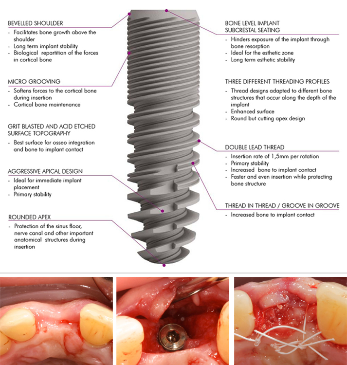
To sum up, the innovative design of the Esthetic Line implant with its sequential thread design, micro-threads to preserve bone at the collar, sophisticated self-cutting and double lead threading preserves bone structure and increases bone to implant contact. This is combined with a SLA surface treatment, a combination of a bevelled shoulder with a platform switching design and a Morse locking conical connection to make it safe and effective in clinical application. The limitation of this study is that the number of cases included is relatively small, only 30 implants of 35 patients were followed up for 12 months. More accurate clinical effect requires more long-term retrospective and prospective clinical observation and research on larger sample size.

References

[1] Albrektsson T,Zarb G,Worthington P,et al. The long – termefficacy of currently used dental implants: a review and proposed criteria of success[J]. The International Journal of Oral & Maxillofacial Implants,1986,1 (1):11-25.

[2] 罗佳,胡秀莲,林野,等. 下后牙区平台转移设计种植体植入深 度对边缘骨水平影响的临床研究[J]. 北京大学学报(医学 版),2012,44 (1):65-69.

[3] 张剑明,郭平川,刘春年,等. 口腔种植修复技术在无牙颌患者 中的应用[J]. 中国口腔种植学杂志,2009,14( 2) : 64 – 65. 1. 陶江丰,陈宁,周芷萱. 口腔种植领域中再生医学的研究进展 [J]. 口腔医学,2012,32(7):443-446.

[4] Kim YK,Kim YJ,Yun PY,et al. Effects of the taper shape,dual- thread and length on the mechanical properties of mini – implants [J]. Angle Orthodontist, 2009, 79(5):908 – 914. 

[5] Hnang HL,Chang CH,Hsu JT,et al. Comparison of implant body designs and threaded designs of dental implants: a 3 – dimensional finite element analysis[J]. In J Oral Maxillofac Implants,2007,22 (4):551-562. 

[6] Bolind K,Johansson CB,Becker W,et al. A descriptive study on re- trieved non – threaded and threaded implant designs[J]. Clinical Oral Implants Research, 2005, 16(4):447 – 455. 

[7] 付丽,周延民. 平台转换结构中肩台变化对种植体 – 骨界面应 力分布的影响[J]. 中国口腔种植学杂志, 2009, 14(2):120.

[8] 郭智舜,郭平川,周贵祥,等. 平台转换种植体肩台宽度对周围 组织的影响[J]. 实用口腔医学杂志, 2013, 29(3):352 – 357.

[9] 宿玉成. 口腔种植学[M]. 第 2 版. 北京: 人民卫生出版社, 2014: 349.

[10] 丁旭,汪大林. 影响种植体及其周围组织应力分布的原因分析 [J]. 口腔颌面修复学杂志, 2008, 9(2):155 – 157.

[11] 董福生, 董玉英,邢汝东,等. 种植体长度对骨界面应力分布影 响 的 三 维 有 限 元 分 析 [ J ] . 中 国 口 腔 种 植 学 杂 志, 2001, 6 (3):106-108.

[12] Nikellis I, Levi A, Nicolopo UC. Immediate loading of 190 endos- seous dental implants: a prospective observational study of 40 pa- tient treatments with up to 2 – year data[J]. The International Journal of Oral & Maxillofacial Implants, 2004, 19(1):116-123.

[13] 宿玉成. 现代口腔种植学[M]. 北京: 人民卫生出版社, 2004: 63-71.

[14] Elias CN, Meirelles L. Improving osseoin tegration of dental implant [J]. Expert Review of Medical Devices,2010,7(2):241-256.

[15] Le Guehennec L,Soueidan A,Layrolle P,et al. Surface treatments of Titanium dental implants for rapid osseointegration[J]. Dental Materials,2007,23(7):844-854.

[16] 刘长虹,杨晓喻. 钛种植体表面特征对种植体周细胞功能的影 响[J]. 中国口腔种植学杂志,2008, 13(2):81-84.

[17] Saivi GG,Gallini G,Lang NP. Early loading (2 or 6 weeks) of sandblasted and acid – etched( SLA) ITI impland in the posterior mandible[J]. Clin Oral Implres,2004,15(1):142-149.

[18] 郭苏伟,赵保东,刘凤芝,等. 平台转换技术对上颌前牙区单枚 种植修复影响的临床观察[J]. 中国口腔种植学杂志,2011,16 (2):120-124.

[19] Otto M. Concept of implant switching [J]. Journal of the South African Dental Association, 2007, 62(2):80.

[20] Lazzara RJ, Porter SS. Platform switching: a new concept in implant dentistry for controlling postrestorative crestal bone levels[J]. The International Journal of Periodontics & Restorative Dentistry, 2006, 26 (1):9 – 1 7 .

[21] 林野. 当代口腔种植学的进展及其临床意义[J]. 口腔颌面外 科杂志, 2006, 16(4):285-290.

[22] 张志勇,王慧明,赖红昌. 口腔颌面种植修复学[M]. 上海: 世 界图书出版公司, 2009: 54.

International Journal of Implant Dentistry – August 2017 – Investigation of peri-implant tissue conditions and peri-implant tissue stability in implants placed with simultaneous augmentation procedure: a 3-year retrospective follow-up analysis of a newly developed bone level implant system

Written by feRKzzCTo0 on . Posted in Century-Gtr, El – Esthetic Line, Henriette Lerner, Robert Sader, Scientific Publications, Shahram Ghanaati, Конвенционална Имплантологија, Регенерација Костију

International Journal of Implant Dentistry • August 2017

Investigation of peri-implant tissue conditions and peri-implant tissue stability in implants placed with simultaneous augmentation procedure: a 3-year retrospective follow-up analysis of a newly developed bone level implant system

Jonas Lorenz University Hospital Frankfurt · Department of oral, maxillofacial and plastic surgery, Henriette Lerner HL DENTCLINIC, Robert A. Sader Goethe-Universität Frankfurt am Main · Center of Stomatology
and Shahram Ghanaati Goethe University of Frankfurt/Main; Universitätsmedizin der Johannes Gutenberg-Universität Mainz · Department for Oral, Craniomaxillofacial and Facial Plastic Surgery; Institute of Pathology

The aim of the present retrospective analysis was to assess peri-implant tissue conditions and document peri-implant tissue stability in C-Tech implants when placed simultaneously with a GBR (Guided Bone Regeneration) augmentation procedure.

A total of 47 implants, which were placed simultaneously with a GBR (Guided Bone Regeneration) procedure with a synthetic bone substitute material in 20 patients, were investigated clinically and radiologically at least 3 years after loading.

The follow-up investigation revealed a survival rate of 100% and only low median rates for probing depths (2.7 mm) and BOP (bleeding on probing) (30%). The mean PES (Pink Esthetic Score) was 10.1 from the maximum value of 14.
No osseous peri-implant defects were obvious, and the mean bone loss was 0.55 mm.

In conclusion, implants placed in combination with a GBR (Guided Bone Regeneration) procedure can achieve long-term stable functionally and esthetically satisfying results for replacing missing teeth in cases of atrophy of the alveolar crest.

References

1. Gurgel BC, Montenegro SC, Dantas PM, Pascoal AL, Lima KC, Calderon PD. Frequency of peri-implant diseases and associated factors. Clin Oral Implants Res. 2016; doi: 10.1111/clr.12944

2. Qian J, Wennerberg A, Albrektsson T. Reasons for marginal bone loss around oral implants. Clin Implant Dent Relat Res. 2012;14(6):792–807.

3. Berglundh T, Lindhe J, Ericsson I, Marinello C, Liljenberg B, Thomsen P. The soft tissue barrier at implants and teeth. Clin Oral Implants Res. 1991;2:81–90.

4. Berglundh T, Lindhe J, Jonsson K, Ericsson I. The topography of the vascular systems in the per iodontal and peri-implant tissues in the dog. J Clin Periodontol. 1999;21:189–93.

5. Moon I, Berglundh T, Abrahamsson I, Linder E, Lindhe J. The barrier between the keratinized mucosa and the dental implant. An experimental study in the dog. J Clin Periodontol. 1999;26:658–63.

6. Lindhe J, Berglundh T. The interface between the mucosa and the implant. Periodontol. 1998;17:47–54.

7. Masaki C, Nakamoto T, Mukaibo T, Kondo Y, Hosokawa R. Strategies for alveolar ridge reconstruction and preservation for implant therapy. J Prosthodont Res. 2015;59(4):220–8.

8. Damien CJ, Parsons JR. Bone graft and bone graft substitutes: areview of current technology and applications. J Appl Biomater.1991;2:187–208.

9. Cordaro L, Torsello F, Miuccio MT, di Torresanto VM, Eliopoulos D. Mandibular bone harvesting for alveolar reconstruction and implant placement: subjective and objective cross-sectional evaluation of donor and recipient site up to 4 y
ears. Clinical Oral Impl Res. 2011;22:1320–6.

10. Canullo L, Penarrocha-Oltra D, Soldini C, Mazzocco F, Penarrocha M, Covani U. Microbiological assessment of the implant-abutment interface in different connections: cross-sectional study after 5 years of functional loading. Clin Oral Implants Res. 2015;26(4):426–34.

11. Misch C. Implant design considerations for the posterior regions of the mouth. Implant Dent. 1999;8(4).

12. SteigengaJ,al-ShammariK,NocitiF,MischC,WangH.Dentalimplant design and its relationship to long-term implant success. Implant Dent. 2001;12(4):306–17.

13. Canullo L, Pace F, Coelho P, Sciubba E, Vozza I. The influence of platform switching on the biomechanical aspects of the implant-abutment system. A three dimensional finite element study. Med Oral Patol Oral Cir Bucal. 2011;16(6):852–6.

14. Lerner H, Lorenz J, Sader R, Ghanaati S. Two-year retrospective study of periimplant health and periimplant bone stability after immediate implant placement of a newly developed bone level implant system—a first report.
EDI Journal (European Association of Dental Implantologists, Teamwork Media); 2017; ahead of print.

15. Ghanaati S, Lorenz J, Obreja K, Choukroun J, Landes C, Sader R. Nanocrystalline hydroxyapatite-base d material already contributes to implant stability after 3 months: a clinical and radiologic 3-year follow-up investigation. In: Journal of Or al Implantology. 2014;40(1):103–9.

16. Lorenz J, Kubesch A, Korzinskas T, Barbeck M, Landes C, Sader R, et al. TRAP-positive multinucleated giant cells are foreign body giant cells rather than osteoclasts: results from a split-mouth study in humans. J Oral Implantol. 2015;41(6):e257–66.

17. Barbeck M, Udeabor S, Lorenz J, Schlee M, Grosse Holthaus M, Raetscho N, et al. High-temperature sintering of xenogeneic bone substitutes leads to increased multinucleated giant cell formation: in vivo and preliminary clinical results. J Oral Implantol. 2015;41(5):e212–22.

18. Barbeck M, Udeabor S, Lorenz J, Kubesch A, Choukroun J, Sader R, et al. Induction of multinucleated giant cells in response to small sized bovine bone substitute (Bio-Oss TM) results in an enhanced early implantation bed vascularization. Ann Maxillofac Surg. 2014;4(2):150–7.

19. Lorenz J, Barbeck M, Sader R, Russe P, Choukroun J, Kirkpatrick CJ, et al. Foreign body giant cell related encapsulation of a synthetic material three years after augmentation. J Oral Implantol. 2016;42(3):273–7.

School of Dental Medicine University of Belgrade, Serbia – November 2015 – Clinical outcomes following horizontal ridge augmentation with bone block grafts

Written by feRKzzCTo0 on . Posted in Century-Gtr, Scientific Publications, Конвенционална Имплантологија, Регенерација Костију

School of Dental Medicine University of Belgrade, Serbia • November 2015

Clinical outcomes following horizontal ridge augmentation with bone block grafts

Assoc Prof Dr Snezana Colic (School of Dental Medicine University of Belgrade, Serbia)

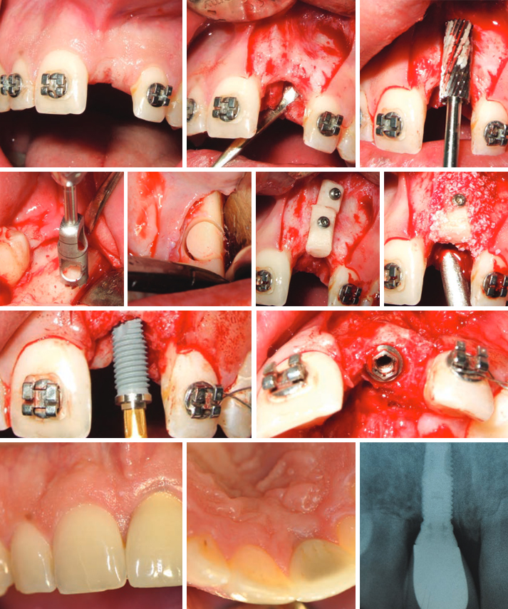
The aim of this study was to analyze clinical outcome of horizontal augmentation with autologous bone block grafts for the reconstruction of narrow edentulous ridge before implant placement. Eighteen partially edentulous patients, presenting insufficient bone width (less than 4mm) in the sites for implant placement were selected.
One or multiple cylindrical block grafts were harvested with trephine burrs from retromolar region and stabilized with titanium miniscrews. Fixed grafts were covered with deproteinised bovine bone mineral granules (DBBM) and collagen membrane (CM). The average amount of bone gain was 3,6 mm. One of the 24 block grafts was lost during the early healing period. Five months later, during the re-entry for implants placement, the gain of ridge width obtained were measured. All implants were placed in correct position and considered successfully integrated during the observation period. Three month after the implant placement prosthetic rehabilitation was started.
The mean follow up after prosthetic load has been 25,4 months.

This technique is reliable means for the correction of narrow edentulous ridge. Close contact between the bone graft and recipient bed surface ensure fast and intense vascularization and subsequent osseointegration of the graft. Adding bovine bone mineral and collagen membrane over bone block minimize resorption during healing.
In all cases all planned implants could be placed in the correct positions.
The mean duration of follow up of the implants was 25,4 months. Prosthodontics rehabilitation involved single crowns or bridges.
Not a single implant was lost during the observation period. All implants were successful both aesthetically and functionally.


References

1. Albrektsson T., Zarb G., Wothington P. & Ericsson A.R. (1986) The long-term efficacy of currently used dental implants: A review and proposed criteria of success. International Journal of Oral and Maxillofacial Implants 1:1-25.

2. Araujo, M.G., Sonohara, M., Hayacibara, R., Car- daropoli, G.
& Lindhe, J. (2002) Lateral ridge augmentation by the use of grafts comprised of autologous bone or a biomaterial. An experiment in the dog. Journal of Clinical Periodontology 29:1122–1131.

3. Buser, D., Dula, K., Belser, U. C., Hirt, H. P. & Berthold, H.
(1995) Localized ridge augmen- tation using guided bone regeneration. II. Surgical procedure in the mandible. International Journal of Periodontics and Restora- tive Dentistry 15,10–29.

4. Buser D., Dula K., Hirt H.P., & Schenk R. (1996) Localized ridge augmentation using autografts and barrier membranes: A clinical study with 40 partially edentulous patients. Journal of Oral and Maxillofacial Surgery 54:420-432

5. Buser, D., Dula, K., Hess, D., Hirt, H.P. & Belser, U.C. (1999) Localized ridge augmentation with autografts and barrier membranes. Periodontology 2000 19:151–163.

6. Buser, D., Ingimarsson, S., Dula, K., Lussi, A., Hirt, H. P. & Belser, U. C. (2002) Long-term stability of osseointegrated implants in aug- mented bone: a 5-year prospective study in partially edentulous patients. International Journal of Periodontics and Restorative Dentistry 22,109–117.

7. Chiapasco M, Romeo E & Vogel G. (1998) Three-dimensional reconstruction of a knife-edge edentulous maxilla by sinus elevation, onlay grafts and sagittal osteotomy of the anterior maxilla: preliminary surgical and prosthetic results. Journal of Oral and Maxillofacial Implants, 13:394-399

8. Chiapasco M., Abati S., Romeo E., Vogel G. (1999) Clinical outcome of autogenous bone blocks or guided bone regeneration with e-PTFE membranes for the reconstruction of narrow edentulous ridges. Clin Oral Impl Res 1999:10:278-288

9. Chiapasco, M., Zaniboni, M. & Boisco, M. (2006) Augmentation procedures for the rehabilitation of deficient edentulous ridges with oral implants. Clin- ical Oral Implants Research 17 (Suppl.):136–159.

10. Cordaro, L., Amade, D. S. & Cordaro, M. (2002) Clinical results of alveolar ridge aug- mentation with mandibular block bone grafts in partially edentulous patients prior to implant placement. Clinical Oral Implants Research 13,103–111.

11. Cordaro, L., Torsello, F., Accorsi Ribeiro, C., Libera- tore, M. & Mirisola di Torresanto, V.M. (2010) Inlay– onlay grafting for three-dimensional reconstruction of the posterior atropic maxilla with mandibular bone. The International Journal of Oral and Maxillofacial Surgery 39:350–357.

12. Cordaro L., Torsello F., Morcavallo S, Mirisola di Torresanto V. (2011) Effect of bovine bone and collagen membranes on healing of mandibular bone blocks: a prospective randomized controlled study. Clin. Oral Impl. Res. 22,2011;1145–1150.

13. Ha ‘mmerle, C.H. & Karring, T. (1998) Guided bone regeneration at oral implant sites. Periodontology 2000 17:151–175.

14. Jensen SS, Aaboe M, Pinholt EM, Hjorting-Hansen E, Melsen F, Ruy- ter IE. Tissue reaction and material characteristics of four bone substitutes. Int J Oral Maxillofac Implants 1996:11:55– 66.

15. Kuboki, Y., Jin, Q., Kikuchi, M., Mamood, J. & Takita, H. (2002) Geometry of artificial ECM: sizes of pores controlling phenotype expression in BMP-induced osteogenesis and chondrogenesis. Connective Tissue Research 43:529–534.

16. Lekholm U & Zarb G.A. (1995) Patient selection and preparation. In: Tissue-integrated prostheses: Osseointegration in clinical dentistry. Pp. 199-209. Chicago: Quintessence

17. Machtei, E.E. (2001) The effect of membrane ex- posure on the outcome of regenerative procedures in humans: a meta-analysis. Journal of Perio- dontology 72:512–516.

18. Misch, C. M. (1997) Comparison of intraoral sites for onlay grafting prior to implant placement. Journal of Oral and
Maxillofacial Implants, 6:767-776

19. Maiorana, C., Beretta, M., Salina, S. & Santoro, F. (2005) Reduction of autogenous bone graft resorption by means of bio-oss coverage: a prospective study. International Journal of Periodontics and Restorative Dentistry 25:19–25.

20. McAllister, B.S. & Haghighat, K. (2007) Bone aug- mentation techniques. Journal of Periodontology 78:377–396.

21. Nkenke, E., Schultze-Mosgau, S., Radespiel-Tro ‘ger, M., Kloss, F. & Neukam, F.W. (2001) Morbidity of harvesting of chin grafts: a prospective study. Clinical Oral Implants Research 12:495–502.

22. Sander, L., Frandsen, E.V., Arnbjerg, D., Warrer, K. & Karring, T. (1994) Effect of local metronidazole application on periodontal healing following guided tissue regeneration. Clinical findings. Journal of Periodontology 65:914–920.

23. Schwarz F., Ferrari D., Balic E., Buser D., Becker J., Sager M. Lateral ridge augmentation using equine- and bovine-derived cancellous bone blocks: a feasibility study in dogs. Clin. Oral Impl. Res. 21,2010;904–912.

24. Simion M., Baldoni M., Rossi P. & Zaffe D. (1994) A comparative study of the effectiveness of a e-PTFE membranes with and without early exposure during healing period. International Journal of Periodontology and Restorative Dentistry 14:167-180

25. Von Arx, T & Buser, D. (2006). Horizontal ridge augmentation using autogenous block grafts and the guided bone regeneration technique with collagen membranes: a clinical study with 42 patients. Clinical Oral Implants Research 17:359–366

Preefooter SER