
3-unit Bridge over Implants using Guided Surgery and One Time Concept
Catarina G. Rodrigues, DDS, MSc – Manuel D. Marques, DDS
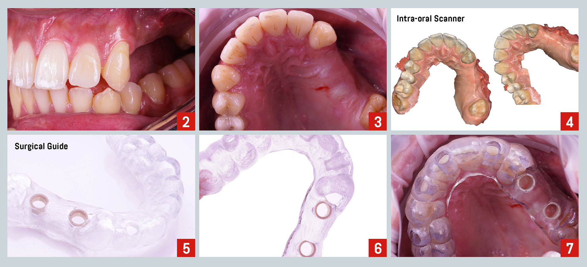
A 30-year-old man presented to a private practice with the chief complaint being “I want to replace the teeth that are missing on my upper jaw”. The clinical and radiographic examination revealed the absence of teeth #24, #25, and #26 (Fig. 2,3). Following a proper diagnosis, the treatment plan proposed was the placement of two implants, followed by the placement of immediate final abutments at the same time of the surgery (One Time Clever Concept, C-Tech Implant). The planned final prosthesis consisted of a 3-unit bridge over implants.
It is described in the literature that the implant–mucosal barrier comprises two components: the junctional epithelium (about 2 mm) and the connective tissue compartment of 1–1.5 mm in height. It was suggested that this attachment serves the purpose of protecting the osseointegration zone. Any disturbance of this zone may affect marginal peri-implant tissues, including peri-implant bone1. In a recent systematic review with meta‐analysis, the authors stated that a repeated abutment disconnection might produce biologic and microbiologic damage to the tissue/abutment/implant complex which considerably increases peri‐implant marginal bone level changes2.
 The One Time concept refers to the placement of the permanent abutment immediately after implant placement, thereby eliminating the need for multiple implant-abutment disconnections. This way, the soft tissue seal around implants is not disrupted and therefore its stability is expected to increase and marginal bone to be preserved.
 In this clinical case, several records of the patient were obtained to digitally plan the surgery: intra-oral photographs, full arch IOS impressions, and CBCT (Fig. 2-4). Once the implant positions were defined, they were translated into the design of the surgical guide (Fig.5-6). The size of the one-time abutments was also planned in the same software as the implants.
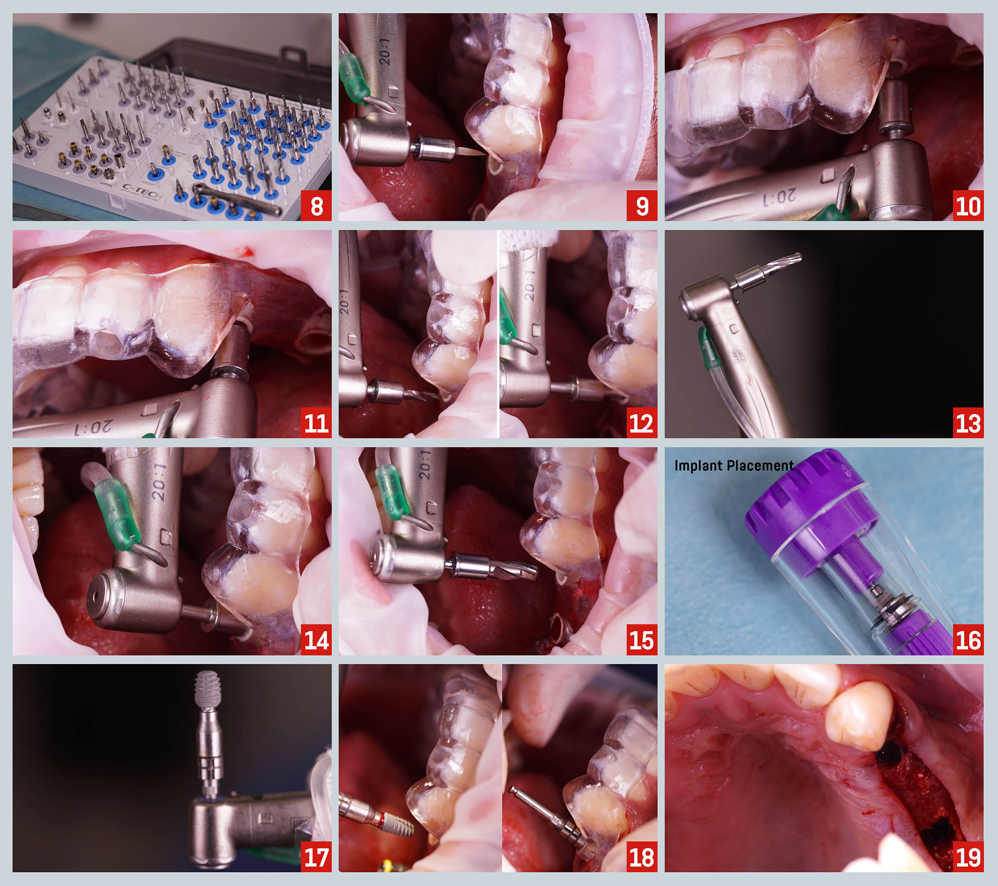
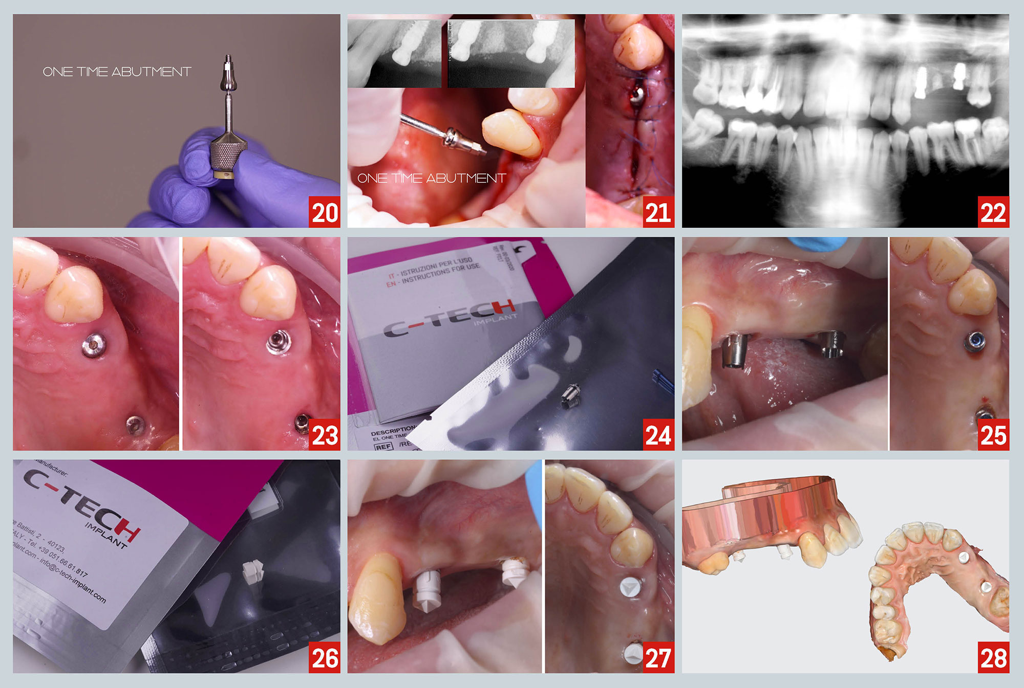
 At the time of the surgery, the stability and adaptation of the guide were checked, followed by guided preparation of the implant sites according to a specific drilling protocol and using C-Tech guided surgery kit (Fig. 7-15). Implants were placed fully guided on 1.4 and 1.6 sites (Fig. 16-19). Then, One Time abutments with 3mm of gingival height were seated on the two implants and fastened with the cover screw which works also as the healing abutment (Fig. 20- 22).
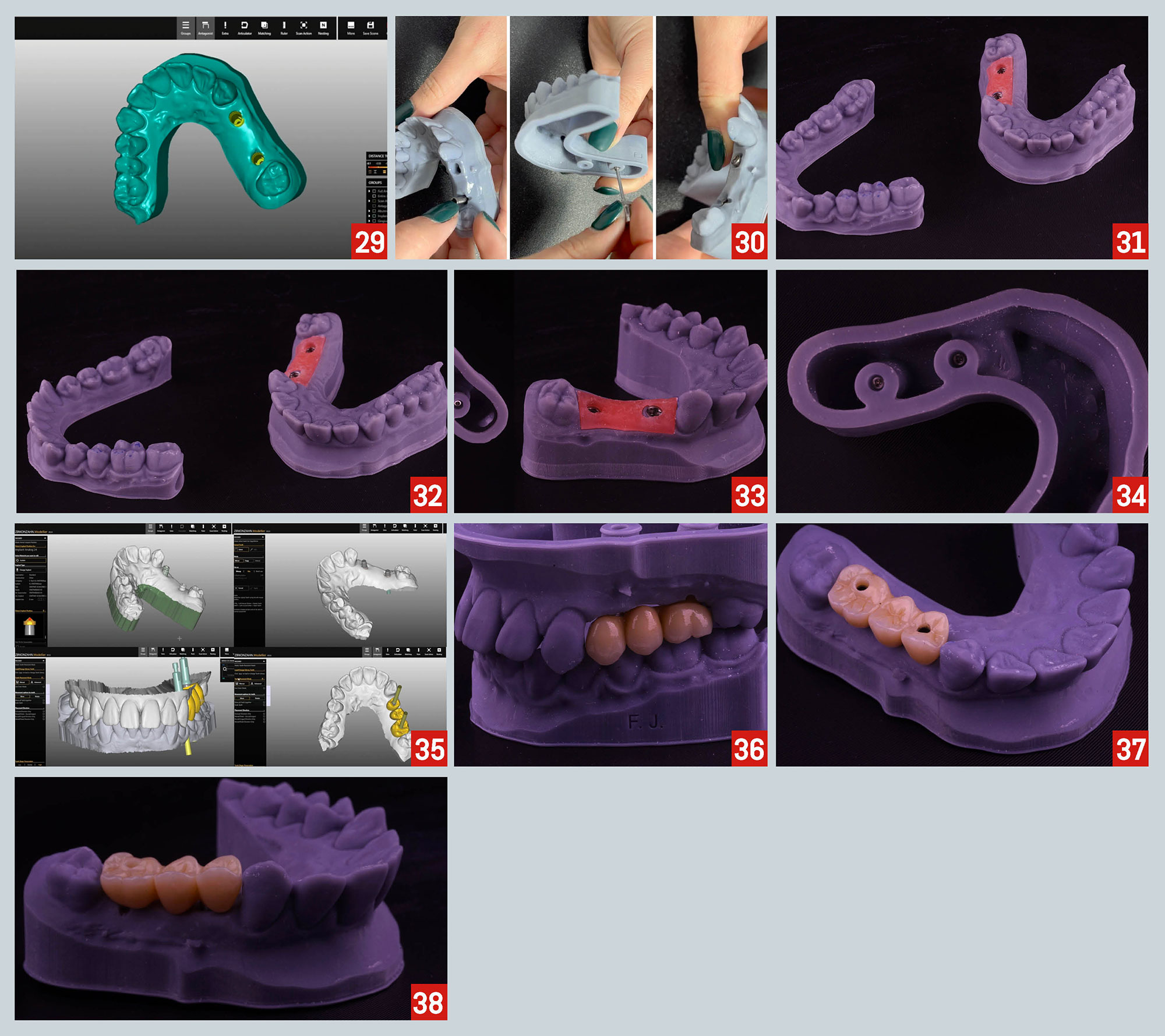
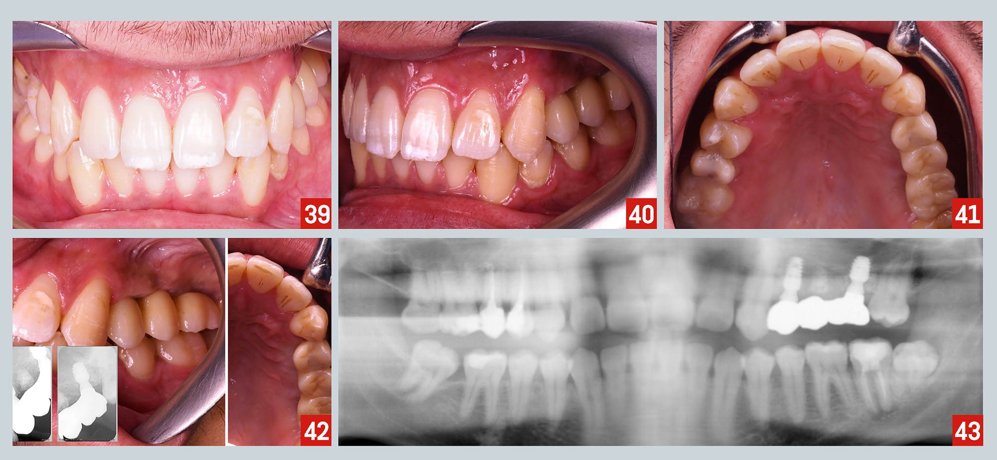 After the osseointegration period, final impressions were taken. First, the cover screw was removed and titanium bases were placed over the One Time abutments. Then, a digital impression was performed using scan caps (Fig. 23-28). The laboratory technician used CAD-CAM technology to obtain a 3D model and to design and mill the final restoration (Fig. 29-38). Finally, the 3-unit bridge over implant was delivered and final records were obtained: intra-oral pictures, periapical and panoramic x-ray (Fig. 39-43).
1-Hamudi, N., Barnea, E., Weinberg, E., Laviv, A., Mijiritsky, E., Matalon, S., Chaushu, L., & Kolerman, R. (2021). The Association of the One-Abutment at One-Time Concept with Marginal Bone Loss around the SLA and Platform Switch and Conical Abutment Implants. Journal of clinical medicine, 11(1), 74.
2-Tallarico M, Caneva M, Meloni SM, Xhanari E, Covani U, Canullo L. Definitive abutments placed at implant insertion and never removed: is it an effective approach? A systematic review and meta‐analysis of randomized controlled trials. J Oral Maxillofac Surg. 2018;76: 316–324.