• Italiano
    • English
    • Deutsch
    • Español
    • Français
    • Русский
    • 中文 (中国)
    • Türkçe
    • Ελληνικά
    • Magyar
    • Српски језик
    • Lietuviškai
    • Bahasa Indonesia
    • Slovenčina

Slide Casi clinici

Συμβατικη Εμφυτευματολογια

Rehabilitation of the jaw by means of immediate implantation and immediate restoration Digital workflow

Written by feRKzzCTo0 on . Posted in Catarina G. Rodrigues, El – Esthetic Line, Gs – Guided Surgery, Manuel D. Marques, Mua, Scientific Publications, Καθοδηγουμενη Χειρουργικη, Συμβατικη Εμφυτευματολογια

Magazine PIP • Practical Implantology and Implant Prosthetics • August 2022 | Issue 4′

Rehabilitation of the jaw by means of immediate implantation and immediate restoration Digital workflow

Catarina G. Rodrigues, DDS, MSc – Manuel D. Marques, DDS – Raquel Bandeira, CDT

The success of any complex dental treatment depends on proper diagnostics and treatment planning. Digital treatment planning, prosthetic backward planning and guided implant surgery can help improve success rates and predictability of complex rehabilitations. The 3D information about the bone anatomy obtained by means of digital volume tomography (DVT) of the patient form the basis for successful treatment planning.

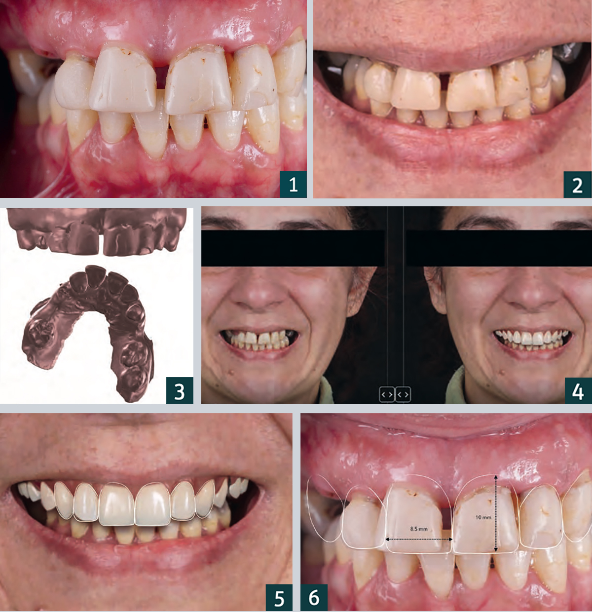
1. Frontal view with centric occlusion preoperative.
2. Preoperative close-up of the patient’s smile.
3. Image data for the preoperative intraoral scan.
4. Virtual simulation of the prosthetic reconstruction using 2D CAD software (Smile Cloud Biometrics; ADN3D Bioetch SRL).
5. Virtual planning of the shape and position of the anterior teeth according to the measurement criteria ..
6. … the red-and-white aesthetic, planned with the 2D Facial Smile design.

PROCEDURE
The DVT data is imported and processed by means of appropriate software in order to plan the implant positions. The planning data is then used to virtually design the drilling template for guided implant placement, which is then 3D printed and used for optimal positioning of the implants. The use of 2D dental CAD software enables the development of a face-related digital smile design. This two-dimensional data can then be imported into 3D CAD software to design 3D mock ups and immediate temporary restorations.

Advantages of immediate implantation
Immediate implantation of implant-supported prostheses is a predictable treatment option in the restoration of an edentulous jaw, as it can contribute to the preservation of the peri-implant soft and hard tissue and enable rapid patient assistance from an aesthetic and functional point of view. Important advantages of using digital technologies for the planning and execution of implant placement as well as for interim restoration are higher accuracy and precision in the fabrication of the overall rehabilitation, low invasiveness and optimal soft tissue shaping by the temporary prosthesis.


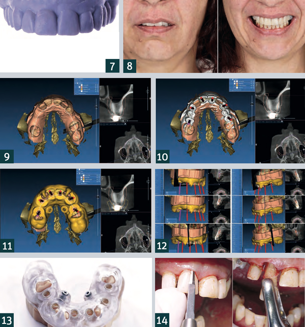
7. Frontal view of the 3D-printed model created from the diagnostic wax-up.
8. Frontal view of the preoperative mock up after transfer to the patient, checking the planning of the 2D facial smile design.
9. Superimposition of the STL data of the preoperative intraoral scan with the DVT data in the planning software (RealGUIDE, 3DIEMME).
10. Digital planning of the implants based on the anatomical situation and the previously planned prosthetic restoration.
11. Once the final implant positions were determined, they were transferred to the surgical template design.
12. Planning of the implant positions.
13. Adjustment of the drilling template on the model.
14. Gentle extraction of the teeth not worth preserving in order to preserve the hard and soft tissue dimensions.

Patient case
The 40-year-old patient presented to our dental practice complaining in particular of difficulty eating and worsened aesthetics in the maxillary anterior region (Figs. 1, 2). The clinical and radiographic examination revealed a bilateral interdental gap in the maxillary posterior region. In addition to a fracture of tooth 13 at gingival level, insufficient amalgam and composite fillings as well as carious defects were diagnosed. There were also increased probing depths and generalised, radiographically visible, horizontal bone loss. After comprehensive diagnostics, it was decided, in consultation with the patient, to extract all of the upper anterior and posterior teeth, and proceed with immediate implantation of a total of eight implants and immediate provisional restoration with screw-retained dentures made of polymethyl methacrylate (PMMA). Intraoral scans (Fig. 3), a DVT and intraoral and extraoral photos (Fig. 4) were taken as part of preoperative diagnosis. In addition, a 2D digital facial smile design was created to enable planning of the position as well as the shape and size of the teeth for the future interim prosthesis (Figs. 5, 6). A digital diagnostic wax-up was then created using 3D CAD software and transferred to a corresponding model using 3D printing (Fig. 7).

Based on the model, a silicone index was created to enable the transfer of the acrylic resin trial restorations to the patient situation and 2D smile planning was undertaken for the patient’s mouth by means of a mock-up (Fig. 8). After adjusting and matching the shape and aesthetics of the anterior teeth, all three-dimensional data (DVT, preoperative intraoral scan and 3D mock-up) were imported into software (Fig. 9) and used to plan the implant position and fabricate the surgical guide (Figs. 10-12). In the patient case under discussion, a mostly fully-navigated implant placement was planned. Special guide sleeves adapted to the implant system were therefore integrated into the surgical template (Fig. 13). The surgical guide was stabilised with tooth support on teeth 16, 12, 22, 26 and 27 and with two palatal bone screws for which two screw channels had been integrated into the surgical guide. Implant preparation was initially fully navigated in the extraction sockets of teeth 13, 11, 21, 23 without opening and in region 15 and 25 with the formation of a mucoperiosteal flap.

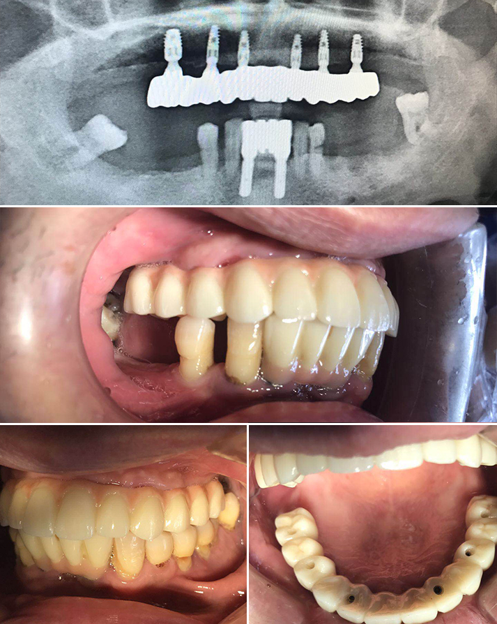
After insertion of the six standard implants in the anterior region, the surgical template was removed, followed by extraction of teeth 16, 12, 22, 26 and 27 (Figs. 14-25). The placement of the two short implants was carried out freehand in region 16 and 26 after removal of the posterior teeth. All implant preparations were carried out according to the manufacturer’s recommended drilling protocol. The provisional prosthesis (Fig. 26) was screwed onto the six anterior implants via abutments and immediately loaded (Fig. 27). A postoperative X-ray was taken (Fig. 28). The patient is very satisfied with her new restoration, both aesthetically and functionally (Figs. 29-31).

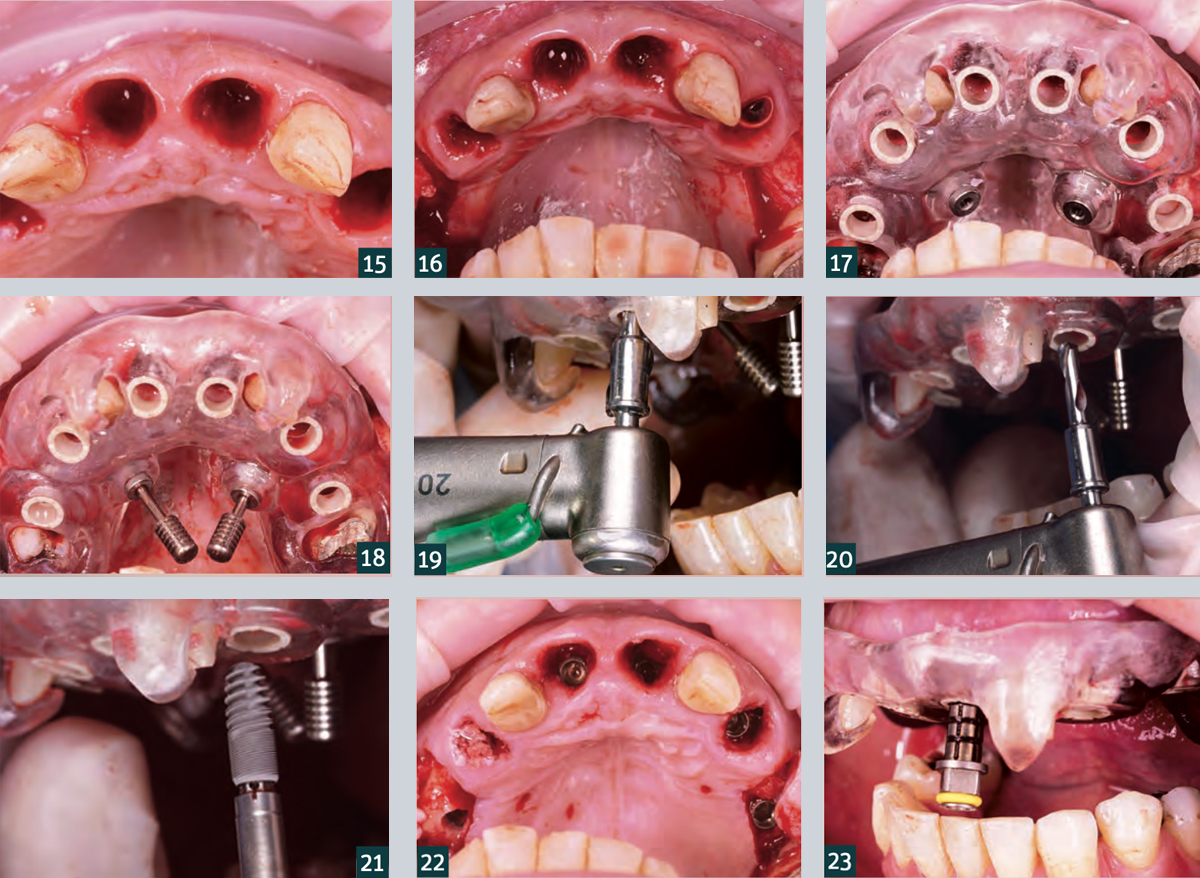
15. Teeth 12 and 22 were initially left in place to stabilise the surgical guide.
16. A full thickness flap was mobilised buccally in the premolar region of the quadrants to increase the soft tissue thickness.
17. Check the intraoral fit and stability of the surgical guide.
18. Additional stabilisation of the surgical guide via palatal fixation screws.
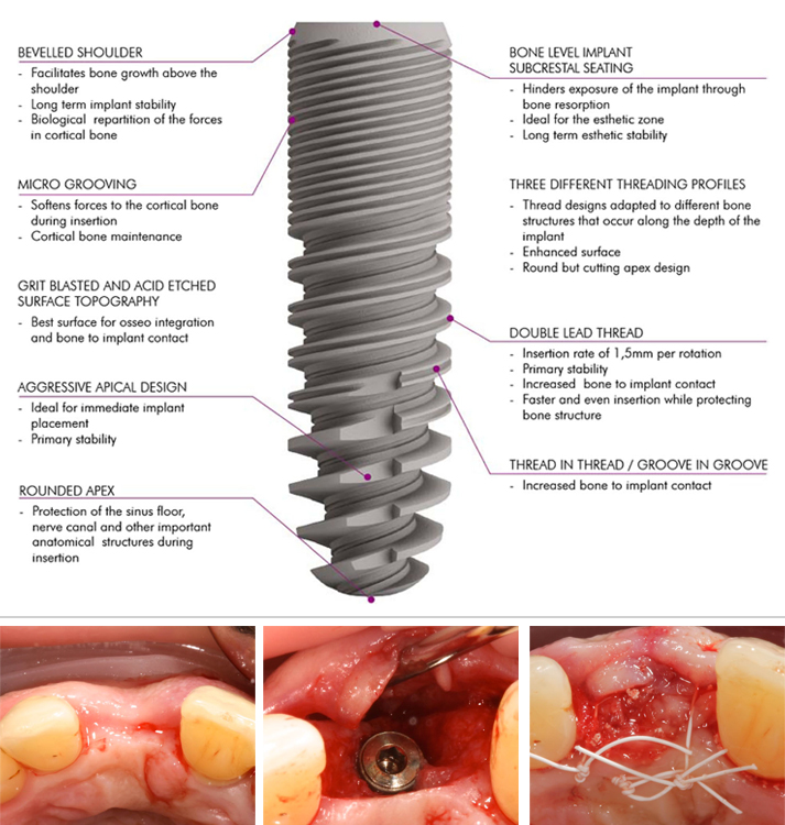
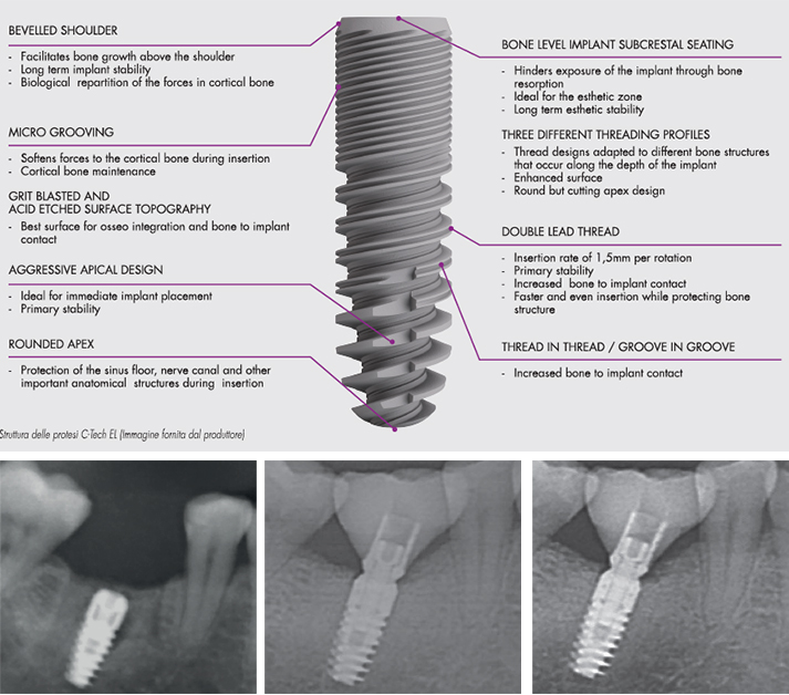
19. Template-guided implant drilling using the C-Guide drill..
20. … for C-Tech implants (C-Tech Implant, Bologna).
21. Template-guided implant placement (Esthetic Line implants, C-Tech Implants).
22. Occlusal view after placement of the six fully-navigated anterior implants.
23. Control: The markings on the transfer post correspond to the surgical guide sleeves.


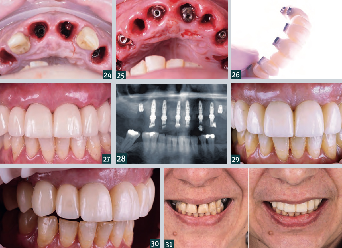
24. Occlusal view of the final implant position in apicocoronal direction (subcrestal positioning of the implants!).
25. Placement of the Multi Unit abutments.
26. Temporary denture made of PMMA.
27. Situation after insertion of the temporary denture with good postoperative soft tissue condition.
28. Radiograph showing postoperative check overview.
29. Intraoral situation one week after the procedure with…
30. … healing without complications and good soft tissue condition.
31. Comparison of the patient situation before (left) and one week after the intervention (right).

Webinar | Treatment of patients with totally edentulous arches: clinical applications of mini implants

Written by feRKzzCTo0 on . Posted in Aldo De Blasi, Scientific Publications, Sd-Mb – Monoblock - Small Diameter, Μικροεμφυτευματα, Συμβατικη Εμφυτευματολογια

Dental Tribune Magazine • Webinar • 11 May 2020

Treatment of patients with totally edentulous arches: clinical applications of mini implants.

by Dr. Aldo De Blasi, expert in dentistry applied to mini implants.

Abstract
Mini implants are a valid alternative to traditional implants to stabilize the rehabilitation of edentulous arches. Patients with full arch prostheses can benefit from this technique which, with a minimally invasive approach, allows to stabilize them with a single operation even in anatomically unfavorable bone conditions. The advantages include a reduced post-operative symptomatology, low-cost therapy and the possibility of using the pre-existing prosthesis. The aim of the seminar is to examine a convenient therapeutic solution, to learn a simple and minimally invasive implant technique and, through the selection of the patient candidate for rehabilitation with mini implants, to discover a new tool for the stabilization of the total prosthesis.

Read the full article on Dental Tribune Magazine
Click here to access the webinar (Italian language)
To access the webinar, you need to register at the following link.


Content of the webinar
This presentation describes the use of mini implants as an anchoring device for a full prosthesis in order to provide greater stability and retention. A simple, fast, convenient and low invasiveness technique is shown, which allows to obtain in a single appointment an effective stabilization of the patient’s removable denture. Where the removable prosthesis is the therapeutic solution to the problem of edentulism, the mini implants can provide additional comfort and well-being. Finally, through the exposure of some clinical cases, the use of these implants with a reduced diameter is shown, it is explained how to select the ideal patient and how to plan the clinical case.

Educational objectives
• Learn a simple and minimally invasive implant technique
• Examin a convenient rehabilitation solution
• Discover a new tool for patient rehabilitation.

Small but powerful. High-quality range with C-Tech system implants.

Written by feRKzzCTo0 on . Posted in El – Esthetic Line, Nd – Narrow Diameter, Scientific Publications, Αναγέννηση Οστών, Καθοδηγουμενη Χειρουργικη, Μικροεμφυτευματα, Συμβατικη Εμφυτευματολογια

Magazine PIP • Practical implantology and implant prosthetics • November 2019

Small but powerful. High-quality range with C-Tech system implants.

Full Smile, under the direction of business owner Lütfü Agic, well-known in the field of dental implantology, advises on a high-quality range with C-Tech system implants, biomaterials such as Bioteck, as well as dental and surgical instruments and tools from devemed. How can such a small unit survive in times of ever greater global players in the field of dentistry and what is the reason for a dentist to look for the partnership, asked pip and talked to Stefan Grümer, M.Sc., who runs together with colleagues a large referral practice on Theaterstrasse in Aachen.

pip: Don’t you kind of lose touch with international developments and trends with such a small partner like Full Smile?
Dr. Grümer: That may well be, regarding these international trends. Here, in the Theaterstraße in Aachen, we are a practice with an impressive range of modern services, especially in the field of restorative and aesthetic dentistry. From 3D diagnostics and computer-based functional diagnostics to laser dentistry, we use state-of-the-art and exceptionally gentle treatment methods that are comfortable for the patient. We are focusing strongly on training programs so that our range of services stays up to date with the latest state of science and research. But we do not have to follow any pseudo innovation. Much more important to us is a certain consistency with our commercial partners, because as well as between us and our patients, the cooperation between us and a medical device manufacturer is a matter of trust. It would bother me to have to adjust to a new contact person every few months, who knows neither me nor my treatment philosophy nor knows what my experiences are and what might actually enrich and interest me. Since 2013 I have been running the first university teaching practice in Germany in cooperation with the RWTH Aachen, an international academy with many foreign and extensive research projects – above all in the field of implantology and laser dentistry. Therefore, we are demanding a certain level of consulting expertise and a portfolio at eye level from our partners.

pip: What impressed you most about the C-Tech Implant System?
Dr. Grümer: As a referral practice we naturally work with different systems. Personally, I especially appreciate the C-Tech Implant System because of its system-related features. It is a fully engineered system that provides different implant diameters and lengths for all common indications – up to special designs such as one-piece mini-implants for narrow spaces, abutments or anchoring of dentures. At C-Tech we experience a very high primary stability and a very easy handling and thus a method which is absolutely suitable for everyday use. Furthermore, the aesthetic demands of our patients have increased significantly. With the EL – Esthetic Line – C-Tech offers a special design for these more demanding challenges. Refinements, like the platform switch, a generous apical threading, the morse taper connection and the concave aesthetic concept create a good stability with excellent shaping of the surrounding tissue. Despite a high prosthetic design freedom, the system is very easy to use, but the prosthetic connection is identical to the other implant diameters of the series. So you can get by with a straightforward instrumentation. I also like a whole new service of Full Smile, which I have never experienced before: As a dentist, I can search for any product via Full Smile and, if I found one, I will only pay the same list price as in the catalogue!

pip: What about service and accessibility – how can such a small company compete with companies that are quite different staffed?
Dr. Grümer: In fact, we sometimes ask ourselves the famous question of how often Mr. Agic has been cloned. He’s really frequently on the move and also visits us regularly to exchange ideas with us or to prepare ourselves for a new technique. However, we always reach someone in the office or receive a callback within a very short time – and also in such a short time our request is accepted, a delivery is initiated or a technical question is answered. In fact, with other companies I sometimes spent a lot more time in some telephone loop with automated speech before I can speak to a human being. With individual consultation, the reliable assurance of the desired delivery times and practical training, Full Smile is a professional partner for us in the field of dental implantology, periodontology and surgery.


Extraction of a Fractured Upper Left Second Premolar, Conventional Implant Placement, with Bone Grafting and Immediate Loading of a Provisional. Final restoration with digital occlusal analysis using T-Scan. – A Case Report

Written by feRKzzCTo0 on . Posted in EL – Esthetic Line, Fabrizia Luongo, Κλινικές περιπτώσεις, Συμβατικη Εμφυτευματολογια

Dr Fabrizia Luongo, DDS, MS, Periodontist, Rome, Italy

Introduction
The evolution of digital dentistry and the development of a digital workflow has concentrated on digital planning with the use of Cone Beam CT scanning as well as using digital restorative tools to combine DICOM (Digital Imaging Communication in Medicine) and .stl (stereolithography) files to virtually plan, place and restore implants before using this plan to treat patients. The resulting benefits are reduced chair time, high precision and predictable aesthetic results often with immediate fixed provisional restorations available at time of surgery and corresponding high levels of patient satisfaction.
Intra-oral scanning to create digital ‘virtual impressions’ is also becoming more prevalent with the information being stored in the .stl file format. This information can be utilised by appropriate CAD/CAM (computer-aided design and computer aided manufacturing) software to design and manufacture a dental restoration (either by milling or 3D printing).
One area that is sometimes overlooked is the use of digital technology in occlusal analysis and adjustment of the restored dental implant. The following case study examines the occlusal management of a conventionally placed implant.

Unconventional Implant Placement Through Impacted Maxillary Canine – A Case Report

Written by feRKzzCTo0 on . Posted in Κλινικές περιπτώσεις, Συμβατικη Εμφυτευματολογια

dr Ivona Bjenjaš, Dental office BGD Osmeh, Belgrade, Serbia

Abstract
We described a case report of a 66 years old patient with asymptomatic impacted maxillary canine free of any surrounding pathology. Extraction of the impacted tooth was avoided because of expected massive bone loss and possible complications. Two C-Tech implants were placed through the impacted tooth in region 13 and 14. Primary stability was achieved on 60N/cm. No postoperative pain, swelling or bleeding was reported by the patient. After four months implants were uncovered and rehabilitated prosthetically with E-max CAD bridge.

Italian passion for German precision

Written by feRKzzCTo0 on . Posted in El – Esthetic Line, Nd – Narrow Diameter, Scientific Publications, Sd-Mb – Monoblock - Small Diameter, Αναγέννηση Οστών, Καθοδηγουμενη Χειρουργικη, Μικροεμφυτευματα, Συμβατικη Εμφυτευματολογια

Magazine PIP • Practical implantology and implant prosthetics • August 2018

Italian passion for German precision

When visiting the impressive manufacturing facility, with 25 CNC machines alone, you will also come across the “Mercedes” of manufacturing technology such as the Swiss Tornos, a five-axis machine, which at C-Tech is purely tasked with the manufacture of mini-implants. Currently they are the largest manufacturer in Europe for this specific type of implant. Mini-implants are popular as a minimally-invasive option for creating an immediate restoration, which is currently a highly sought-after alternative for fast and good functional care of older patients. An impressive neighbour is the Willemin Macodell (the Rolls-Royce of CNC machines), which can be fitted with 48 different CNC cutters. It can change tools in 1.4 seconds and can be used in any conceivable axis, including for the manufacture of angled mini-abutments. C-Tech was one of the first companies to move over to cooling all computer-controlled machine tools using water-miscible bio-lubricants. The plant-based oil is odour-free, improves machining and even has a wider range of applications than mineral oil products. “And even though of course we run extensive cleaning processes for the finished parts, it’s still one risk factor less”, comments Lütfü Agic with respect to the recently published independent surface study by the University of Cologne, where the C-Tech implant surfaces were tested as an example, in testing applied to very few dental implant surfaces on the dental market.

Erfolgsfaktoren für die Behandlung mit MiniImplantaten und ihre Bedeutung für die Praxis – Praktische Implantologie und Implantatprothetik | pip 4 | 2010

Written by feRKzzCTo0 on . Posted in Henriette Lerner, Scientific Publications, Sd-Mb – Monoblock - Small Diameter, Μικροεμφυτευματα, Συμβατικη Εμφυτευματολογια

Praktische Implantologie und Implantatprothetik | pip 4 | 2010

Erfolgsfaktoren für die Behandlung mit MiniImplantaten und ihre Bedeutung für die Praxis. Prospektive Untersuchung von Patientenfällen über ein Jahr

Literatur bei den Verfassern: Henriette Lerner, Ady Palti

Der klinische Erfolg von Mini-Implantaten hängt von verschiedenen Parametern ab, die auch untereinander in Beziehung stehen. In der vorliegenden Studie wurde untersucht, ob eine Korrelation zwischen dem Durchmesser der inserierten Implantate und der Primärstabilität existiert. Dabei wurden zudem unterschiedliche, durch den jeweiligen Patientenfall vorgegebene Knochendichten berücksichtigt. Außerdem erfolgte eine Dokumentation der Osseointegration und der Tiefe etwaiger perioimplantärer Taschen über einen Zeitraum von einem Jahr post implantationem. Darüber hinaus wurden der Einfluss des Implantatdurchmessers und der Art des Implantataufbaus auf die Erfolgsrate ermittelt.

Schlussfolgerung

Aufgrund der im Rahmen der vorliegenden Untersuchung ermittelten Ergebnisse darf bei einer Verwendung von Mini-Implantaten zur Prothesenstabilisierung mit einer Erfolgsrate ähnlich wie bei klassischen Implantationen gerechnet werden. Da der Erfolg mit der Primärstabilität korreliert, lässt er sich nach deren Bestimmung mit der Drehmomentratsche direkt im Anschluss an die Insertion bereits gut abschätzen. Liegen die ermittelten Zahlen im grenzwertigen Bereich (≈ 35 Ncm), so sollte im Zweifelsfalle eine weiche Unterfütterung vorgenommen werden. Je nach individuellem Fall ist auch zu prüfen, ob zur besseren Stabilisierung ein weiteres Mini-Implantat inseriert werden kann. Hat man die Wahl, so sollte eher ein etwas größeres gewählt werden, d.h. statt des 1,8-mm-Implantats lieber das 2,1er bzw. statt des 2,4-mm-Implantats lieber das MDI Hybrid mit 2,9 mm Durchmesser.

Es versteht sich von selbst, dass eng beieinander liegende Recall-Termine angeraten sind, insbesondere um die planmäßige Osseointegration zeitnah verfolgen zu können. Sie ist keinesfalls nach sechs Monaten abgeschlossen, sondern erfährt in den darauffolgenden sechs Monaten in der Regel noch einmal eine signifikante Verbesserung.

Dr. medic.stom. Henriette Lerner

1990 Studium der Zahnmedizin (Universität für Medizin und Pharmazie „Victor Babes“ Temeschburg).
1990-1993 Oralchirurgische Weiterbildung an der Akademie für Zahnärztliche Fortbildung Karlsruhe.
1995 Training in Goldman School of Dental Implantology/Boston, Massachussets.
1998 Spezialist DGZI.
2004 Expert Implantologie der DGOI.
2006-2007 Spezialisierung “Dento-alveoläre Chirurgie” Dr. medic.stom. Henriette Lerner (Universität “Carol Davila” Bukarest).
2006 Praxis im Videnti Zentrum für Implantologie und Ästhetik, Baden Baden.
Mitglied in: DGOI; ICOI; EAO; ASA DGÄZ; DGZMK; BDO; EFOSS.
Nationale und Internationale Referententätigkeit über Ästhetik in der Implantologie, Minimal Invasive Implantology, Curriculum Implantologie, fortgeschrittene Augmentationstechniken.

A one year follow up examining bone level preservation utilising platform switching implants – Ningxia Med J Dec. 2017

Written by feRKzzCTo0 on . Posted in Century-Gtr, Scientific Publications, Αναγέννηση Οστών, Συμβατικη Εμφυτευματολογια

Ningxia Med J, Dec. 2017, Vol 39, No. 12

A one year follow up examining bone level preservation utilising platform switching implants

Jianlin Chen, Yuanjie Cao, Lili Shan, Yan Li, Li Ma, Na Wang, Xiuyu Wu Author’s
Unit: Department of Stomatology, Lingwu People’s Hospital, Ningxia

Abstract

Objective The aim of this research is to observe and evaluate the clinical application of the Esthetic Line [EL] implant system (C-Tech, Bologna, Italy). The EL implant has a surface topography created by sandblasting and acid etching (SLA) and a Morse locking conical connection.

Methods 35 patients with one or more missing teeth were selected and a total of 60 EL implants were placed using either a one stage (non-submerged) placement protocol or a two stage (submerged) protocol. Where applicable, second stage surgery was undertaken 2 ~ 4 months post implantation. Subsequent to this the EL implants were permanently restored over a period of 2 ~ 4 weeks. Clinical examination and imaging analysis were undertaken to evaluate clinical success.

Results All 60 implants osseointegrated successfully, and at the one year follow up there was a 100% retention rate and no adverse reactions in the host. Mesial and distal bone heights were recorded on the day of surgery as well as at the fitting of the permanent restoration and after the implants had been functionally loaded for 12 months. Mesial bone heights were (0. 35 ± 0. 49) mm, (0. 18 ± 0. 44) mm and (0. 25 ± 0. 36) mm respectively. Distal bone heights were (0. 20 ± 0. 42) mm, (0. 08 ± 0. 45) mm and (0. 15 ± 0. 38) mm. In the first year of implant functional load, the total absorption of the mesial bone was (- 0. 11 ± 0. 38) mm and the distance was (- 0. 07 ± 0. 31) mm; There was no significant difference in blood indexes between the preoperative and postoperative three months (P < 0. 05).

Conclusion The design of the EL implant incorporating a combination of a parallel walled section with an apical taper, a beveled shoulder, platform switching, a Morse locking conical connection combined with an SLA treated surface and a sophisticated double threaded morphological design all contribute to successful treatment.

Key words Italy; C – Tech Esthetic Line [EL] implant; implant design; surface treatment; Morse-locking.

In recent years, the evolution of implant techniques combined with developments in implant design and restorative techniques has resulted in greater acceptance of implant treatment by the majority of patients. The Stomatology department of our hospital has adopted the Esthetic Line [EL] implant system (C-Tech, Bologna, Italy) for use in our clinic. A study into the clinical results of 60 EL implants placed in 35 patients was undertaken.  

Conclusions

To sum up, the innovative design of the Esthetic Line implant with its sequential thread design, micro-threads to preserve bone at the collar, sophisticated self-cutting and double lead threading preserves bone structure and increases bone to implant contact. This is combined with a SLA surface treatment, a combination of a bevelled shoulder with a platform switching design and a Morse locking conical connection to make it safe and effective in clinical application. The limitation of this study is that the number of cases included is relatively small, only 30 implants of 35 patients were followed up for 12 months. More accurate clinical effect requires more long-term retrospective and prospective clinical observation and research on larger sample size.

References

[1] Albrektsson T,Zarb G,Worthington P,et al. The long – termefficacy of currently used dental implants: a review and proposed criteria of success[J]. The International Journal of Oral & Maxillofacial Implants,1986,1 (1):11-25.

[2] 罗佳,胡秀莲,林野,等. 下后牙区平台转移设计种植体植入深 度对边缘骨水平影响的临床研究[J]. 北京大学学报(医学 版),2012,44 (1):65-69.

[3] 张剑明,郭平川,刘春年,等. 口腔种植修复技术在无牙颌患者 中的应用[J]. 中国口腔种植学杂志,2009,14( 2) : 64 – 65. 1. 陶江丰,陈宁,周芷萱. 口腔种植领域中再生医学的研究进展 [J]. 口腔医学,2012,32(7):443-446.

[4] Kim YK,Kim YJ,Yun PY,et al. Effects of the taper shape,dual- thread and length on the mechanical properties of mini – implants [J]. Angle Orthodontist, 2009, 79(5):908 – 914. 

[5] Hnang HL,Chang CH,Hsu JT,et al. Comparison of implant body designs and threaded designs of dental implants: a 3 – dimensional finite element analysis[J]. In J Oral Maxillofac Implants,2007,22 (4):551-562. 

[6] Bolind K,Johansson CB,Becker W,et al. A descriptive study on re- trieved non – threaded and threaded implant designs[J]. Clinical Oral Implants Research, 2005, 16(4):447 – 455. 

[7] 付丽,周延民. 平台转换结构中肩台变化对种植体 – 骨界面应 力分布的影响[J]. 中国口腔种植学杂志, 2009, 14(2):120.

[8] 郭智舜,郭平川,周贵祥,等. 平台转换种植体肩台宽度对周围 组织的影响[J]. 实用口腔医学杂志, 2013, 29(3):352 – 357.

[9] 宿玉成. 口腔种植学[M]. 第 2 版. 北京: 人民卫生出版社, 2014: 349.

[10] 丁旭,汪大林. 影响种植体及其周围组织应力分布的原因分析 [J]. 口腔颌面修复学杂志, 2008, 9(2):155 – 157.

[11] 董福生, 董玉英,邢汝东,等. 种植体长度对骨界面应力分布影 响 的 三 维 有 限 元 分 析 [ J ] . 中 国 口 腔 种 植 学 杂 志, 2001, 6 (3):106-108.

[12] Nikellis I, Levi A, Nicolopo UC. Immediate loading of 190 endos- seous dental implants: a prospective observational study of 40 pa- tient treatments with up to 2 – year data[J]. The International Journal of Oral & Maxillofacial Implants, 2004, 19(1):116-123.

[13] 宿玉成. 现代口腔种植学[M]. 北京: 人民卫生出版社, 2004: 63-71.

[14] Elias CN, Meirelles L. Improving osseoin tegration of dental implant [J]. Expert Review of Medical Devices,2010,7(2):241-256.

[15] Le Guehennec L,Soueidan A,Layrolle P,et al. Surface treatments of Titanium dental implants for rapid osseointegration[J]. Dental Materials,2007,23(7):844-854.

[16] 刘长虹,杨晓喻. 钛种植体表面特征对种植体周细胞功能的影 响[J]. 中国口腔种植学杂志,2008, 13(2):81-84.

[17] Saivi GG,Gallini G,Lang NP. Early loading (2 or 6 weeks) of sandblasted and acid – etched( SLA) ITI impland in the posterior mandible[J]. Clin Oral Implres,2004,15(1):142-149.

[18] 郭苏伟,赵保东,刘凤芝,等. 平台转换技术对上颌前牙区单枚 种植修复影响的临床观察[J]. 中国口腔种植学杂志,2011,16 (2):120-124.

[19] Otto M. Concept of implant switching [J]. Journal of the South African Dental Association, 2007, 62(2):80.

[20] Lazzara RJ, Porter SS. Platform switching: a new concept in implant dentistry for controlling postrestorative crestal bone levels[J]. The International Journal of Periodontics & Restorative Dentistry, 2006, 26 (1):9 – 1 7 .

[21] 林野. 当代口腔种植学的进展及其临床意义[J]. 口腔颌面外 科杂志, 2006, 16(4):285-290.

[22] 张志勇,王慧明,赖红昌. 口腔颌面种植修复学[M]. 上海: 世 界图书出版公司, 2009: 54.

Aspects of oral morphology as decision factors in mini-implant supported overdenture – Romanian Journal of Morphology and Embryology • 2010, 51(2):309–314

Written by feRKzzCTo0 on . Posted in Henriette Lerner, Scientific Publications, Sd-Mb – Monoblock - Small Diameter, Μικροεμφυτευματα, Συμβατικη Εμφυτευματολογια

Romanian Journal of Morphology and Embryology • 2010, 51(2):309–314

Aspects of oral morphology as decision factors in mini-implant supported overdenture

Elena Preoteasa, Marina MeleŞcanu-imre, Cristina Teodora Preoteasa Department of Oral Diagnosis and Ergonomics “Carol Davila” University of Medicine and Pharmacy of Bucharest, Romania, Mihaela Marin Department of Prosthodontics, Henriette Lerner Private practice, Baden Baden, Germany

Abstract

Evaluation of some morphological oral aspects perceived as decision factors in complete edentulism treatment by mini-implants overdenture. Patients, Material and Methods: An observational study was conducted on a sample of 24 patients (average age of 61 years), through clinical and imagistic methods. The variables taken into consideration were: age, gender, alveolar mucosa status, bone offer, miniimplants characteristics, insertion torque and loading type. Results: 117 mini-implants were applied. Conclusions: Mini-implant supported overdenture can be an alternative to conventional denture and conventional implant overdenture. Its advantages derives from implants’ characteristics (smaller diameter, variable length, O-ring retention system), which adapts better to the particular edentulous conditions. Insertion technique implies less surgical trauma. Choosing implants’ size, number, topography, and the loading method have a great variety, depending on anatomical feature (bone offer, mucosa and relationship with the nearby anatomical structures), functional features and patients’ wishes.

Conclusions

Mini-dental implant supported overdenture can be in complete edentulous patients a treatment alternative to both conventional dentures and conventional implant retained overdenture.

The advantages of this type of treatment derives from the characteristics of this type of implant (small diameter, variable length, O-ring retention system), which adapts better to the particular morphological conditions present in full edentulous patients. Also, the implants insertion requires less surgical trauma, this being a beneficial aspect in the context of usually poor general status.

Proceeding and achieving the treatment requires needs careful evaluation through clinical, imagistic and laboratory methods. Frequently there are identified some unfavorable conditions for implant insertion and a high degree of treatment difficulty. These issues (morphological and functional features, related to general health status, age, gender, etc.) must be linked to implants characteristics, in order to decide the particular treatment features, from surgical and prosthetic point of view. The length, diameter, number, topography, loading method of implants can present a large variety, depending on quantitative bone offer (ridge width and bone height), quality (bone density), functional features and patient’s wishes.

This type of treatment has a lower cost compared to conventional implant supported overdenture, by the lower cost of mini-implants, and also due to the use, in general, just of the panoramic radiography as imagistic method (computed tomography is an adjuvant method, but not essential in most cases). Also by eliminating some surgical intervention, we eliminate also their costs. On the other hand, the benefits related to an improved stability, better functionality and adaptation are quickly noticed by the patient and increases their level of satisfaction.

Due to the relatively simple technique, less traumatic, but with benefits that are quickly perceived, the mini-implant supported overdenture may be the elective treatment alternative for complete edentulous patients.

References

[1] PREOTEASA E, BĂNCESCU G, LONESCU E, BĂNCESCU A, DONCIU D, Epidemiologic aspects of the totally edentulous mouth. (1) General aspects, Bacteriol Virusol Parazitol Epidemiol, 2004, 49(3–4):115–120.

[2] PREOTEASA E, LONESCU E, BĂNCESCU G, BĂNCESCU A, PREOTEASA CT, Epidemiologic aspects of the edentulous mouth (II). General and local features of the totally edentulous mouth, Bacteriol Virusol Parazitol Epidemiol, 2005, 50(1–2):27–34.

[3] MELESCANU M, PREOTEASA E, Mandibular panoramic indexes predictors of skeletal osteoporosis for implant therapy, Curr Health Sci J, 2009, 35(4):291–296.

[4] FRIEDLANDER AH, The physiology, medical management and oral implications of menopause, J Am Dent Assoc, 2002, 133(1):73–81.

[5] ROBERTS WE, SIMMONS KE, GARETTO LP, DECASTRO RA, Bone physiology and metabolism in dental implantology: risk factors for osteoporosis and other metabolic bone diseases, Implant Dent, 1992, 1(1):11–21.

[6] LERNER H, Minimal invasive implantology with small diameter implants, Implant Pract, 2009, 2(1):30–35.

[7] ***, IMTEC Corporation official website, retrieved December 13, 2009, http://www.imtec.com/europe/.

[8] GRIFFITTS TM, COLLINS CP, COLLINS PC, Mini dental implants: an adjunct for retention, stability, and comfort for the edentulous patient, Oral Surg Oral Med Oral Pathol Oral Radiol Endod, 2005, 100(5):e81–e84.

[9] BALKIN BE, STEFLIK DE, NAVAL F, Mini-dental implant insertion with the auto-advance technique for ongoing applications, J Oral Implantol, 2001, 27(1):32–37.

[10] FROUM SJ, SIMON H, CHO SC, ELIAN N, ROHRER MD, TARNOW DP, Histological evaluation of bone-implant contact of immediately loaded transitional implants after 6 to 27 months, Int J Oral Maxillofac Implants, 2005, 20(1):54–60.

[11] MORNEBURG TR, PRÖSCHEL PA, Success rates of microimplants in edentulous patients with residual ridge resorption, Int J Oral Maxillofac Implants, 2008, 23(2):270–276.

[12] LABARRE EE, AHLSTROM RH, NOBLE WH, Narrow diameter implants for mandibular denture retention, J Calif Dent Assoc, 2008, 36(4):283–286.

[13] ORDOÑEZ A, EASTMOND V, Mini-dental implants – they’re here to stay, Postgraduate Dentist Caribbean, 3(1):7–12.

[14] SHATKIN TE, SHATKIN S, OPPENHEIMER AJ, Mini dental implants for the general dentist: a novel technical approach for small-diameter implant placement, Compendium, 2003, 24:26–34. st [15] MISCH CE, Dental implant prosthetics, 1 edition, Mosby, St. Louis, 2005, 130–141.

International Journal of Implant Dentistry – August 2017 – Investigation of peri-implant tissue conditions and peri-implant tissue stability in implants placed with simultaneous augmentation procedure: a 3-year retrospective follow-up analysis of a newly developed bone level implant system

Written by feRKzzCTo0 on . Posted in Century-Gtr, El – Esthetic Line, Henriette Lerner, Robert Sader, Scientific Publications, Shahram Ghanaati, Αναγέννηση Οστών, Συμβατικη Εμφυτευματολογια

International Journal of Implant Dentistry • August 2017

Investigation of peri-implant tissue conditions and peri-implant tissue stability in implants placed with simultaneous augmentation procedure: a 3-year retrospective follow-up analysis of a newly developed bone level implant system

Jonas Lorenz University Hospital Frankfurt · Department of oral, maxillofacial and plastic surgery, Henriette Lerner HL DENTCLINIC, Robert A. Sader Goethe-Universität Frankfurt am Main · Center of Stomatology
and Shahram Ghanaati Goethe University of Frankfurt/Main; Universitätsmedizin der Johannes Gutenberg-Universität Mainz · Department for Oral, Craniomaxillofacial and Facial Plastic Surgery; Institute of Pathology

The aim of the present retrospective analysis was to assess peri-implant tissue conditions and document peri-implant tissue stability in C-Tech implants when placed simultaneously with a GBR (Guided Bone Regeneration) augmentation procedure.

A total of 47 implants, which were placed simultaneously with a GBR (Guided Bone Regeneration) procedure with a synthetic bone substitute material in 20 patients, were investigated clinically and radiologically at least 3 years after loading.

The follow-up investigation revealed a survival rate of 100% and only low median rates for probing depths (2.7 mm) and BOP (bleeding on probing) (30%). The mean PES (Pink Esthetic Score) was 10.1 from the maximum value of 14.
No osseous peri-implant defects were obvious, and the mean bone loss was 0.55 mm.

In conclusion, implants placed in combination with a GBR (Guided Bone Regeneration) procedure can achieve long-term stable functionally and esthetically satisfying results for replacing missing teeth in cases of atrophy of the alveolar crest.

References

1. Gurgel BC, Montenegro SC, Dantas PM, Pascoal AL, Lima KC, Calderon PD. Frequency of peri-implant diseases and associated factors. Clin Oral Implants Res. 2016; doi: 10.1111/clr.12944

2. Qian J, Wennerberg A, Albrektsson T. Reasons for marginal bone loss around oral implants. Clin Implant Dent Relat Res. 2012;14(6):792–807.

3. Berglundh T, Lindhe J, Ericsson I, Marinello C, Liljenberg B, Thomsen P. The soft tissue barrier at implants and teeth. Clin Oral Implants Res. 1991;2:81–90.

4. Berglundh T, Lindhe J, Jonsson K, Ericsson I. The topography of the vascular systems in the per iodontal and peri-implant tissues in the dog. J Clin Periodontol. 1999;21:189–93.

5. Moon I, Berglundh T, Abrahamsson I, Linder E, Lindhe J. The barrier between the keratinized mucosa and the dental implant. An experimental study in the dog. J Clin Periodontol. 1999;26:658–63.

6. Lindhe J, Berglundh T. The interface between the mucosa and the implant. Periodontol. 1998;17:47–54.

7. Masaki C, Nakamoto T, Mukaibo T, Kondo Y, Hosokawa R. Strategies for alveolar ridge reconstruction and preservation for implant therapy. J Prosthodont Res. 2015;59(4):220–8.

8. Damien CJ, Parsons JR. Bone graft and bone graft substitutes: areview of current technology and applications. J Appl Biomater.1991;2:187–208.

9. Cordaro L, Torsello F, Miuccio MT, di Torresanto VM, Eliopoulos D. Mandibular bone harvesting for alveolar reconstruction and implant placement: subjective and objective cross-sectional evaluation of donor and recipient site up to 4 y
ears. Clinical Oral Impl Res. 2011;22:1320–6.

10. Canullo L, Penarrocha-Oltra D, Soldini C, Mazzocco F, Penarrocha M, Covani U. Microbiological assessment of the implant-abutment interface in different connections: cross-sectional study after 5 years of functional loading. Clin Oral Implants Res. 2015;26(4):426–34.

11. Misch C. Implant design considerations for the posterior regions of the mouth. Implant Dent. 1999;8(4).

12. SteigengaJ,al-ShammariK,NocitiF,MischC,WangH.Dentalimplant design and its relationship to long-term implant success. Implant Dent. 2001;12(4):306–17.

13. Canullo L, Pace F, Coelho P, Sciubba E, Vozza I. The influence of platform switching on the biomechanical aspects of the implant-abutment system. A three dimensional finite element study. Med Oral Patol Oral Cir Bucal. 2011;16(6):852–6.

14. Lerner H, Lorenz J, Sader R, Ghanaati S. Two-year retrospective study of periimplant health and periimplant bone stability after immediate implant placement of a newly developed bone level implant system—a first report.
EDI Journal (European Association of Dental Implantologists, Teamwork Media); 2017; ahead of print.

15. Ghanaati S, Lorenz J, Obreja K, Choukroun J, Landes C, Sader R. Nanocrystalline hydroxyapatite-base d material already contributes to implant stability after 3 months: a clinical and radiologic 3-year follow-up investigation. In: Journal of Or al Implantology. 2014;40(1):103–9.

16. Lorenz J, Kubesch A, Korzinskas T, Barbeck M, Landes C, Sader R, et al. TRAP-positive multinucleated giant cells are foreign body giant cells rather than osteoclasts: results from a split-mouth study in humans. J Oral Implantol. 2015;41(6):e257–66.

17. Barbeck M, Udeabor S, Lorenz J, Schlee M, Grosse Holthaus M, Raetscho N, et al. High-temperature sintering of xenogeneic bone substitutes leads to increased multinucleated giant cell formation: in vivo and preliminary clinical results. J Oral Implantol. 2015;41(5):e212–22.

18. Barbeck M, Udeabor S, Lorenz J, Kubesch A, Choukroun J, Sader R, et al. Induction of multinucleated giant cells in response to small sized bovine bone substitute (Bio-Oss TM) results in an enhanced early implantation bed vascularization. Ann Maxillofac Surg. 2014;4(2):150–7.

19. Lorenz J, Barbeck M, Sader R, Russe P, Choukroun J, Kirkpatrick CJ, et al. Foreign body giant cell related encapsulation of a synthetic material three years after augmentation. J Oral Implantol. 2016;42(3):273–7.

EDI Journal – December 2016 – First report of a two-year retrospective study with a newly developed bone-level implant system Peri-implant health and peri-implant bone stability after immediate implant placement

Written by feRKzzCTo0 on . Posted in El – Esthetic Line, Henriette Lerner, Robert Sader, Scientific Publications, Shahram Ghanaati, Συμβατικη Εμφυτευματολογια

International Journal of Implant Dentistry • December 2016

First report of a two-year retrospective study with a newly developed bone-level implant system Peri-implant health and peri-implant bone stability after immediate implant placement

DR-Medic Stom. Henriette Lerner HL DENTCLINIC, Dr. Jonas Lorenz University Hospital Frankfurt · Department of oral, maxillofacial and plastic surgery, Professor Robert A. Sader Goethe-Universität Frankfurt am Main · Center of Stomatology and Dr. Shahram Ghanaati Goethe University of Frankfurt/Main; Universitätsmedizin der Johannes Gutenberg-Universität Mainz · Department for Oral, Craniomaxillofacial and Facial Plastic Surgery; Institute of Pathology

Dental implants have become a reliable and predictable treatment modality to replace missing teeth and retain dentures in edentulous patients. They can restore the oral health, form, function, mastication, articulation and aesthetics of the stomatognathic system with multi-year success rates of more than 90 per cent for implants in fully edentulous [1,2] or partially edentulous patients [3-6]. Variations in implant success have been found dependent upon surgical technique, loading protocol, implant localisation and bone quality –for example, lower success rates have been reported for maxillary implants than for mandibular implants [7,8]

In the past few decades, research on dental implants has led to a broad modification of the surgical and prosthetic protocols.
For the surface of dental implants there is a clear consensus regarding the superiority of roughened/micro-textured surfaces.

Other ways to increase the implant surface include the thread design, implant length and implant diameter. The implant design should incorporate features that best transform tensile and shear forces during mastication and minimize undesirable force components.
Also essential for the long-term stability of peri-implant bone tissue and an aesthetically and functionally sufficient dental implant is the stability of the implant/abutment connection, to prevent implant fractures and screw loosening and to keep the peri-implant bone level stable.
A space or micro-gap between the implant and abutment is unavoidable with a two-piece design; however, a smaller micro-gap can also sometimes be found in designs with platform switching and Morse-tapered conical connectors, used to transfer the micro-gap facing the implant axis and reduce micro-movement. This can reduce the propulsion of sulcus fluid and, consequently, crestal bone loss, even with implants inserted below the alveolar crest (subcrestally).

The aim of the present retrospective study was to describe the clinical and radiological results of a new implant system with a grit-blasted and acid-etched surface topography and a Morse-locking conical implant/abutment connection. Implants were inserted in fresh and intact extraction sockets of maxillary and mandibular non-salvageable teeth and were followed up clinically and radiologically after a mean loading time of two years. Special emphasis was placed on the maintenance of peri-implant health and the stability of peri-implant bone level.


References

1. Albrektsson T, Dahl E, Enbom L, Engevall S, Engquist B, et al. (1988) Osseointegrated oral implants. A Swedish multicenter study of 8139 consecutively inserted nobelpharma implants. J Periodontol 59(5):287-296.

2. Spiekermann H, Jansen VK, Richter EJ (1995) A 10-year follow-up study of IMZ and TPS implants in the edentulous mandible using bar-retained overdentures. Int J Oral
Maxillofac Implants 10(2):231-243.

3. Nevins M, Langer B (1993) The successful application of osseointegrated implants to the posterior jaw: a long-term retrospective study. Int J Oral Maxillofac Implants 8(4):428-432.

4. Henry PJ, Laney WR, Jemt T, Harris D, Krogh PH, et al. (1996) Osseointegrated implants for single-tooth replacement: a prospective 5-year multicenter study. Int J Oral Maxillofac Implants 11(4):450-455.

5. Schmitt A, Zarb GA (1993) The longitudinal clinical effectiveness of osseointegrated dental implants for single-tooth replacement. Int J Prosthodont 6(2):197-202.

6. Fugazzotto PA, Gulbransen HJ, Wheeler SL, Lindsay JA (1993) The use of IMZ osseointegrated implants in partially and completely edentulous patients: success and failure rates of 2,023 implant cylinders up to 60+ months in function. Int J Oral Maxillofac Implants 8(6):617-621.

7. Misch CE (1990) Density of bone: effect on treatment plans, surgical approach, healing, and progressive boen loading. Int J Oral Implantol 6(2):23-31.

8. Albrektsson T, Lekholm U (1989) Osseointegration: current state of the art. Dent Clin North Am 33 (4):537-554.

9. Adell R, Lekholm U, Brånemark PI (1985) Surgical procedures. In: Brånemark PI, Zarb G, Albrektsson T (Eds.), Tissue Integrated Prostheses: Surgical Procedures, Quintessence Publishing Co, Chicago, USA, pp. 223-225.

10. Kohal RJ, LaRosa M, Patrick D, Hürzeler MB, Caffesse RG (1999) Clinical and histologic evaluation of submerged and nonsubmerged hydroxyapatite-coated im-plants: a preliminary study in dogs. Int J Oral Maxillofac Implants 14(6):824-834.

11. Evian CI, Kessler L, Axler J (1997) One-stage surgery with a nonsubmerged implant system. Compend Contin Educ Dent 18(11):1091-1094,1096-1098.

12. Ericsson I, Nilner K, Klinge B, Glantz PO (1996) Radio-graphical and histological characteristics of submerged and nonsubmerged titanium implants. An experimental study in the Labrador dog. Clin Oral Implants Res 7(1):20-26.

13. Chrcanovic BR, Albrektsson T, Wennerberg A (2015) Dental implants inserted in fresh extraction sockets versus healed sites: a systematic review and meta-analysis. J Dent 43(1):16-41.

14. Weiss CM, Weiss A, Rosenlicht J (2001) Root form implants. Treatment of total mandibular edentulism diagnosed for an overdenture. In: Weiss CM, Weiss A, (Eds.), Principles and Practice of Implant Dentistry, Mosby, St Louis, USA, pp. 147-168.

15. Fickl S, Zuhr O, Wachtel H, Stappert CF, Stein JM, et al. (2008) Dimensional changes of the alveolar ridge contour after different socket preservation techniques. J Clin Peri-odontol 35(10):906-913.

16. Vignoletti F, Matesanz P, Rodrigo D, Figuero E, Martin C, et al. (2012) Surgical protocols for ridge preservation after tooth extraction. A systematic review. Clin Oral Implants Res 23(5):22-38.

17. Lekovic V, Camargo PM, Klokkevold PR, Weinlaender M, Kenney EB, et al. (1998) Preservation of alveolar bone in extraction sockets using bioabsorbable membranes. J Periodontol 69(9):1044-1049.

18. Becker W, Becker B, Polizzi G, Bergstrom C (1994) Autogenous bone grafting of defects adjacent to implants placed into immediate extraction sockets in patients: a prospective study. Int J Oral Maxillofac Implants 9(4):389-396.

19. Artzi Z, Tal H, Dayan D (2001) Porous bovine bone mineral in healing of human extraction sockets: 2. Histo-chemical observations at 9 months. J Periodontol 72(2):152-159.

20. Iasella JM, Greenwell H, Miller RL, Hill M, Drisko C (2003) Ridge preservation with freeze-dried bone allograft and a collagen membrane compared to extraction alone for implant site development: a clinical and histologic study in humans. J Periodontol 74(7):990-999.

21. Araújo MG, Lindhe J (2005) Dimensional ridge alterations following tooth extraction. An experimental study in the dog. J Clin Periodontol 32(2):212-218.

22. Evans CD, Chen ST (2008) Esthetic outcomes of immediate implant placements. Clin Oral Implants Res 19(1):73-80.

23. Chen ST, Darby IB, Reynolds EC, Clement JG (2009) immediate implant placement postextraction without flap elevation. J Periodontol 80(1):163-172.

24. Lang NP, Pun L, Lau KY, Li KY, Wong MC (2012) A systematic review on survival and success rates of implants placed immediately into fresh extraction sockets after at least 1 year. Clin Oral Implants Res 23(5):39-66.

25. Pagni G, Pellegrini G, Giannobile WV, Rasperini G (2012) Postextraction alveolar ridge preservation: biological basis and treatments. Int J Dent 2012:151030.

26. Tavarez RR, Calixto AM, Maia Filho EM, Bandeca MC, Firoozmand LM, et al. (2014) Atraumatic extraction, implant placement and immediate provisionalization. J Contemp Dent Pract 15(4):513-517.

27. Misch CE (1999) Implant design considerations for the posterior regions of the mouth. Implant Dent 8(4):376-386.

28. Binon PP (2000) Implants and components: entering the new millennium. Int J Oral Maxillofac Implants 15(1):76-94.

29. Trisi P, Rao W, Rebaudi A (1999) A histometric comparison of smooth and rough titanium implants in human low-density jawbone. Int J Oral Maxillofac Implants 14(5):689-698.

30. Steigenga JT, Shammari KF, Nociti FH, Misch CE, Wang HL (2003) Dental implant design and its relationship to long-term implant success. Implant Dent 12(4):306-317.

31. Niznick G (2000) Achieving Osseointegration in soft bone: The search for improved results. Oral Health 90:27-32.

32. O’Sullivan D, Sennerby L, Meredith N (2000) Measurements comparing the initial stability of five designs of dental implants: a human cadaver study. Clin Implant Dent Relat Res 2(2):85-92.

33. Sykaras N, Iacopino AM, Marker VA, Triplett RG, Woody RD (2000) Implant materials, designs, and surface topographies: their effect on osseointegration. A literature review. Int J Oral Maxillofac Implants 15(5):675-690.

34. Canullo L, Penarrocha OD, Soldini C, Mazzocco F, Penarrocha M (2015) Microbiological assessment of the implant-abutment interface in different connections: cross-sectional study after 5 years of functional loading. Clin Oral Implants Res 26(4):426-434.

35. Pozzi A, Tallarico M, Moy PK (2014) Three-year post-loading results of a randomised, con-trolled, split-mouth trial comparing implants with different prosthetic interfaces and design in partially posterior edentulous mandibles. Eur J Oral Implantol 7(1):47-61.

36. Ghanaati S, Lorenz J, Obreja K, Choukroun J, Landes C, et al. (2014) Nanocrystalline hydroxyl apatite-based material already contributes to implant stability after 3 months: a clinical and radiologic 3-year followup investigation. J Oral Implantol 40(1):103-109.

37. Ghanaati S, Barbeck M, Lorenz J, Stuebinger S, Seitz O (2013) Synthetic bone substitute material comparable with xeno-geneic material for bone tissue regeneration in oral cancer patients: First and preliminary histological, histomorpho-metrical and clinical results. Ann Maxillofac Surg 3(2):126-138.

38. Sailer I, Zembic A, Jung RE, Siegenthaler D, Holderegger C (2009) Randomized controlled clinical trial of customized zirconia and titanium implant abutments for canine and posterior single-tooth implant reconstructions: preliminary results at 1 year of function. Clinical Oral Implants Research 20(3):219-225.

39. Brägger U, Bürgin WB, Hämmerle CH, Lang NP (1997) Associations between clinical parameters assessed around implants and teeth. Clinical Oral Implants Research 8(5):412-421.

40. Berglundh T, Lindhe J, Ericsson I, Marinello CP, Liljenberg B, et al. (1991) The soft tissue barrier at implants and teeth. Clinical Oral Implants Research 2(2):81-90.

41. Berglundh T, Lindhe J, Jonsson K, Ericsson I (1994) The topography of the vascular systems in the periodontal and peri-implant tissues in the dog. Journal of Clinical Periodontology 21(3):189-193.

42. Moon IS, Berglundh T, Abrahamsson I, Linder E, Lindhe J (1999) The barrier between the keratinized mucosa and the dental implant. An experimental study in the dog. Journal of Clinical Periodontology 26(10):658-663.

43. Lindhe J, Berglundh T (1998) The interface between the mucosa and the implant. Periodontology 2000 17: 47-54.

44. Rieder D, Eggert J, Krafft T, Weber HP, Wichmann MG (2014) Impact of placement and restoration timing on single-implant esthetic outcome-a randomized clinical trial. Clin Oral Implants Res 27(2):e80-e86.

45. Romanos GE, Aydin E, Locher K, Nentwig GH (2014): Immediate vs. delayed loading in the posterior mandible: a split-mouth study with up to 15 years of follow-up. Clin Oral Implants Res 27(2):e74-e79.

46. Wagenberg B, Froum SJ (2014) Long-Term Bone Stability around 312 Rough-Surfaced Immediately Placed Implants with 2-12-Year Follow-Up. Clin Implant Dent Relat Res 17(4):658-666.

47. Javed F, Ahmed HB, Crespi R, Romanos GE (2013) Role of primary stability for successful osseointegration of dental implants: Factors of influence and evaluation. Interv Med Appl Sci 5(4):162-167.

48. Calvo GJL, Gomez MG, Aguilar SA, Mate SVJE, Abboud M (2014) Bone remodeling at implants with different configurations and placed immediately at different depth into extraction sockets. Experimental study in dogs. Clin Oral Implants Res 26(5):507-515.

49. Javed F, Almas K, Crespi R, Romanos GE (2011) Implant surface morphology and primary stability: is there a connection? Implant Dent 20(1):40-46.

50. Berberi A, Tehini G, Rifai K, Bou NEF, El ZN (2014) In vitro evaluation of leakage at implant-abutment connection of three implant systems having the same prosthetic interface using rhodamine B. Int J Dent 2014:351263.

Dentista Moderno, Italy – October 2016 – Follow-up after two years to check the health of peri-implant tissues and bone stability of a number of cases with immediate post-extraction insertion of a recently developed bone implant system

Written by feRKzzCTo0 on . Posted in El – Esthetic Line, Henriette Lerner, Robert Sader, Scientific Publications, Shahram Ghanaati, Συμβατικη Εμφυτευματολογια

Dentista Moderno, Italy • October 2016

Follow-up after two years to check the health of peri-implant tissues and bone stability of a number of cases with immediate post-extraction insertion of a recently developed bone implant system.First report

Jonas Lorenz University Hospital Frankfurt · Department of oral, maxillofacial and plastic surgery, Henriette Lerner HL DENTCLINIC, Robert A. Sader Goethe-Universität Frankfurt am Main · Center of Stomatology
and Shahram Ghanaati Goethe University of Frankfurt/Main; Universitätsmedizin der Johannes Gutenberg-Universität Mainz · Department for Oral, Craniomaxillofacial and Facial Plastic Surgery; Institute of Pathology

Introduction
Dental implants have become a reliable and predictable treatment method in dentistry to replace the tooth and prosthesis in case of edentulism. Therefore the oral health, shape, operation, chewing, articulation and aesthetics of the stomatognathic apparatus can be restored with a multi-year success rate of more than 90% in the case of implants set in completely or partially edentulous patients.
For the success of an implant in the long term it is necessary to meet some specific technical and constructive requirements. An additional factor that is essential for the long-term stability of peri-implant bone tissue and to ensure an aesthetically and functionally adequate dental implant is the stability of the implant-abutment connection, as it is inevitable that a space or micro-gap will be created between the implant and the abutment. Nevertheless, there will be a smaller micro-gap if the design is equipped with a cone Morse connection and platform switching, which transfer the micro-gap to the front of the implant axis and reduce micro-motion. Therefore, the pumping of sulcular fluid and, consequently, crestal bone loss can be reduced even when the implant is inserted under the crest (subcrestal).

The purpose of the series of cases presented is to describe, for the first time, the clinical and radiological results after two years of immediate setting of 50 implants with a new sandblasted and acid-etched surface implant system and cone Morse connection.

During the observation period which averaged two years, none of the implants presented failures or acute or peri-implant infections. All implants had a sufficient amount of keratinised peri-implant soft tissue, reduced probing depths (2.25 mm on average) and good BOP (34%). After about two years of use, peri-implant bone level was stable and with an average bone loss of 0.83 mm.

With an average observation period of two years, the implant considered with immediate setting on the bone, rough surface and conical connection was shown to preserve the health status of the soft and hard peri-implant tissues. The parameters analysed are the same or better than comparable studies in the international literature.


References

1. Albrektsson, I; Dahl, E.; Enbom, L; Engevall, S.; Engquist, B.; Eriksson, A. R. et al. (1988): Osseointegrated oral implants. A Swedish multicenter study of 8139 consecutively inserted Nobelpharma implants. In: J. Periodontal 59 (5):287-296.

2. Spiekermann, H.; Jansen, V. K.; Richter, E. J. (1995): A 10-year follow-up study of IMZ and TPS implants in the edentulous mandible using bar-retained overdentures. In: Int J Oral Maxillofac Implants 10 (2):231 -243.

3. Nevins, M.; Longer, B. (1993): The successful application of osseointegrated implants to the posterior jaw: a long-term retrospective study. In: Int J Oral Maxillofac Implants 8 (4):428-432.

4. Henry, P. J.; Laney, W. R.; Jemt, T.; Harris, D.; Krogh, P. H.; Polizzi, G. et al. (1996): Osseointegrated implants for single-tooth replacement: a prospective 5-year multicenter study. In: Int J Oral Maxillofac Implants 11 (4):450-455.

5. Schmitt, A.; Zarb, G. A. (1993): The longitudinal clinical effectiveness of osseointegrated dental implants for single-tooth replacement. In: Int J Prosthodont 6 (2):197-202.

6. Fugazzotto, P. A.; Gulbransen, H. J.; Wheeler S. L.; Lindsay, J. A. (1993): The use of IMZ osseointegrated implants in partially and completely edentulous patients: success and failure rates of 2,023 implant cylinders up to 60+ months in function. In: Int J Oral Maxillofac Implants 8 (6):617-621.

7. Misch, C. E. (1990): Density of bone: effect on treatment plans, surgical approach, healing, and progressive boen loading. In: Int J Oral Impiantai 6 (2):23-31.

8. Albrektsson, T.; Lekholm, U. (1989): Osseointegration: current state of the art. In: Dent. Clin. North Am 33 (4):537-554.

9. Misch, C. E. (1999): Implant design considerations for the posterior regions of the mouth. In: Implant Dent 8 (4):376-386.

10. Binon, P. P. (2000): Implants and components: entering the new millennium. In: Int J Oral Maxillofac Implants 15 (1):76-94.

11. Trisi, R; Rao, W; Rebaudi, A. (1999): A histometric comparison of smooth and rough titanium implants in human low-density jawbone. In: Int J Oral Maxillofac Implants 14 (5):689-698.

12. Steigenga, Jennifer T.; al-Shammari, Khalaf R; Nociti, Francisco H.; Misch, Carl E.; Wang, Horn-Lay (2003): Dental implant design and its relationship to long-term implant success. In: Implant Dent 12 (4):306-317.

13. Niznick, G. (2000): Achieving Osseointegration in soft bone: The search for improved results. In: Oral Health, 2000;90:27-32.

14. O’Sullivan, D.; Sennerby, L; Meredith, N. (2000): Measurements comparing the initial stability of five designs of dental implants: a human cadaver study. In: Clin Implant Dent Relat Res 2 (2):85-92.

15. Sykaras, N.; Iacopino, A. M.; Marker, V. A.; Triplett, R. G.; Woody, R. D. (2000): Implant materials, designs, and surface topographies: their effect on osseointegration. A literature review. In: Int J Oral Maxillofac Implants 15 (5):675-690.

16. Canullo, Luigi; Penarrocha-Oltra, David; Soldini, Claudio; Mazzocco, Fabio; Penarrocha, Maria; Covani, Ugo (2015): Microbiological assessment of the implant-abutment interface in different connections: cross-sectional study after 5 years of functional loading. In: Clin Oral Implants Res 26 (4):426-434

17. Pozzi, Alessandro; Tallarico, Marco; Moy. Peter K. (2014): Three-year post-loading results of a randomised, controlled, split-mouth trial comparing implants with different prosthetic interfaces and design in partially posterior edentulous mandibles. In: Eur J Oral Impiantai 7 (1):47-61.

18. Ghanaati, Shahram; Lorenz, Jonas; Obreja, Karina; Choukroun, Joseph; Landes, Constantin; Sader, Robert A. (2014): Nanocrystalline hydroxyapatite-based material already contributes to implant stability after 3 months: a clinical and radiologic 3-year follow-up investigation. In: J Oral Impiantai 40 (1):103-109.

19. Ghanaati, Shahram; Barbeck, Mike; Lorenz, Jonas; Stuebinger, Stefan; Seitz, Oliver; Landes, Constantin et al. (2013): Synthetic bone substitute material comparable with xenogeneic material for bone tissue regeneration in oral cancer patients: First and preliminary histological, histomorphometrical and clinical results. In: Ann Maxillofac Sura 3 (2):126-138.

20. Sailer, I; Zembic, A; Jung, R; Siegenthaler, D; Holderegger, C; Hàmmerle, C. (2009): Randomized controlled clinical trial or customized zirconio and titanium implant abutments for canine and posterior single-tooth implant reconstructions: preliminary results at 1 year of function. In: Clinical Oral Implants Research; 20:219-225.

21. Bràgger, U; Burgin, W; Hàmmerle, C; Lang N. (1997): Associations between clinical parameters assessed around implants and teeth. In: Clinical Oral Implants Research; 8:412-421.

22. Tavarez, Rudys Rodolfo Jesus de; Calixto, Amanda Martins; Maia Filho, Etevaldo Matos; Bandeca, Matheus Coelho; Firoozmand, Leily Macedo; Gomes, Mario Gilson Nina; Malheiros, Adriana Santos (2014): Atraumatic extraction, implant placement and immediate provisionalization. In: J Contemp Dent Pract 15 (4):513-517.

23. Rieder, Dominik; Eggert, Jochen; Krafft, Tim; Weber, Hans-Peter; Wichmann, Manfred G.; Heckmann, Siegfried M. (2014): Impact of placement and restoration timing on single-implant esthetic outcome – a randomized clinical trial. In: Clin Oral Implants Res.

24. Romanos, Georgios E.; Aydin, Erhan; Locher, Kathrin; Nentwig, Georg-Hubertus (2014): Immediate vs. delayed loading in the posterior mandible: a split-mouth study with up to 15 years of follow-up. In: Clin Oral Implants Res.

25. Berberi, Antoine; Tehini, Georges; Rifai, Khaldoun; Bou Nasser Eddine, Farah; El Zein, Nabil; Badran, Bassam; Akl, Haidar (2014): In vitro evaluation of leakage at implant-abutment connection of three implant systems having the same prosthetic interface using rhodamine B. In: Int J Dent 2014:351263.

Xian Feng Dental Magazine – July 2016 – The Past and Present State of the Art in Dental Implants

Written by feRKzzCTo0 on . Posted in El – Esthetic Line, Scientific Publications, Συμβατικη Εμφυτευματολογια

Xian Feng Dental Magazine • July 2016

The Past and Present State of the Art in Dental Implants

Dr Wei

Abstract
Implants have existed for thousands of years, and titanium implants have existed since 1965. There have been many developments in titanium implants in the last 50 years. The culmination of all the developments and the present state of the art in dental implants is demonstrated by the EL (Esthetic Line) dental implant by C-TECH. The EL implant superior performance is due to its main characteristics: Platform switching, Morse lock, tapered, conical connection as well as a bevelled shoulder and agressive main body threading. The Morse lock conical connection brings substantial benefits: connection strength, protection against bacterial infiltration of the gap, hindrance of screw loosening and consequent prosthetic failure. The generous main body threading provides excellent bone to implant contact as well as excellent performance in soft bone.

Preefooter GR