Dr Fabrizia Luongo, DDS, MS, Periodontist, Rome, Italy
Introduction
The use of Cone Beam Computerised Tomography (CBCT) scanning is becoming more commonplace in today’s implant dentistry. These scans combined with increasingly sophisticated software technology has led to greater accuracy in digital planning and guided surgery and is gradually being adopted across general practice. Furthermore, the restorative planning can be digitised and combined with the CBCT data in appropriate planning software.
In conjunction with these technology advances, dental implant design has evolved to incorporate the latest concepts of biomechanical design, which include sophisticated thread profiling, platform switching and a Morse locking taper on the implant/abutment interface.
The following case details brings these advances in implants and digital technologies together.
Case Study
A 64-year-old female patient presented with an endodontically failed upper left pre-molar. Figures 14a & 14b Provisional bridge in place.
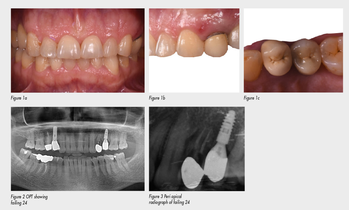
Figures 1a, b & c initial presentation of patient showing endodontically failed 24.
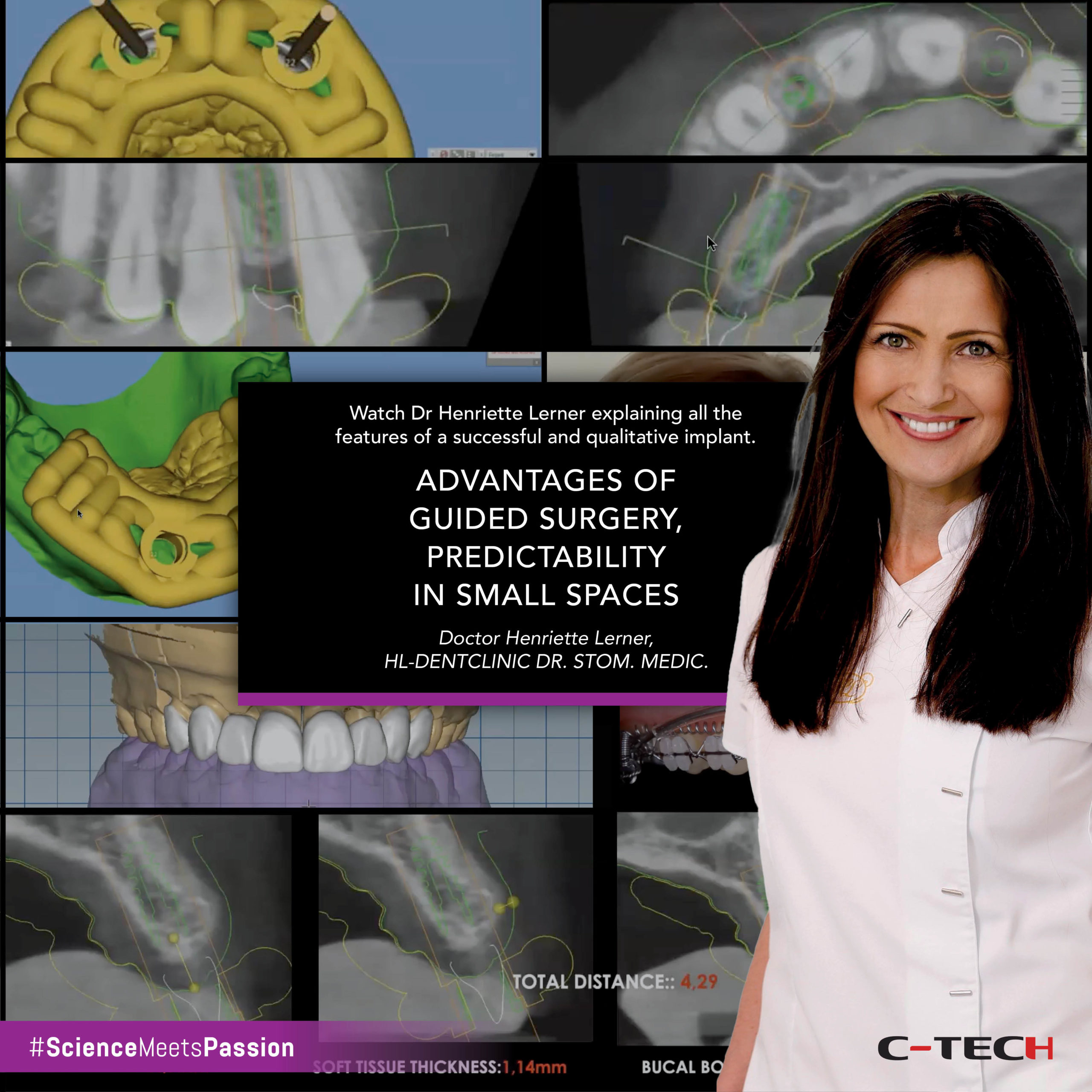
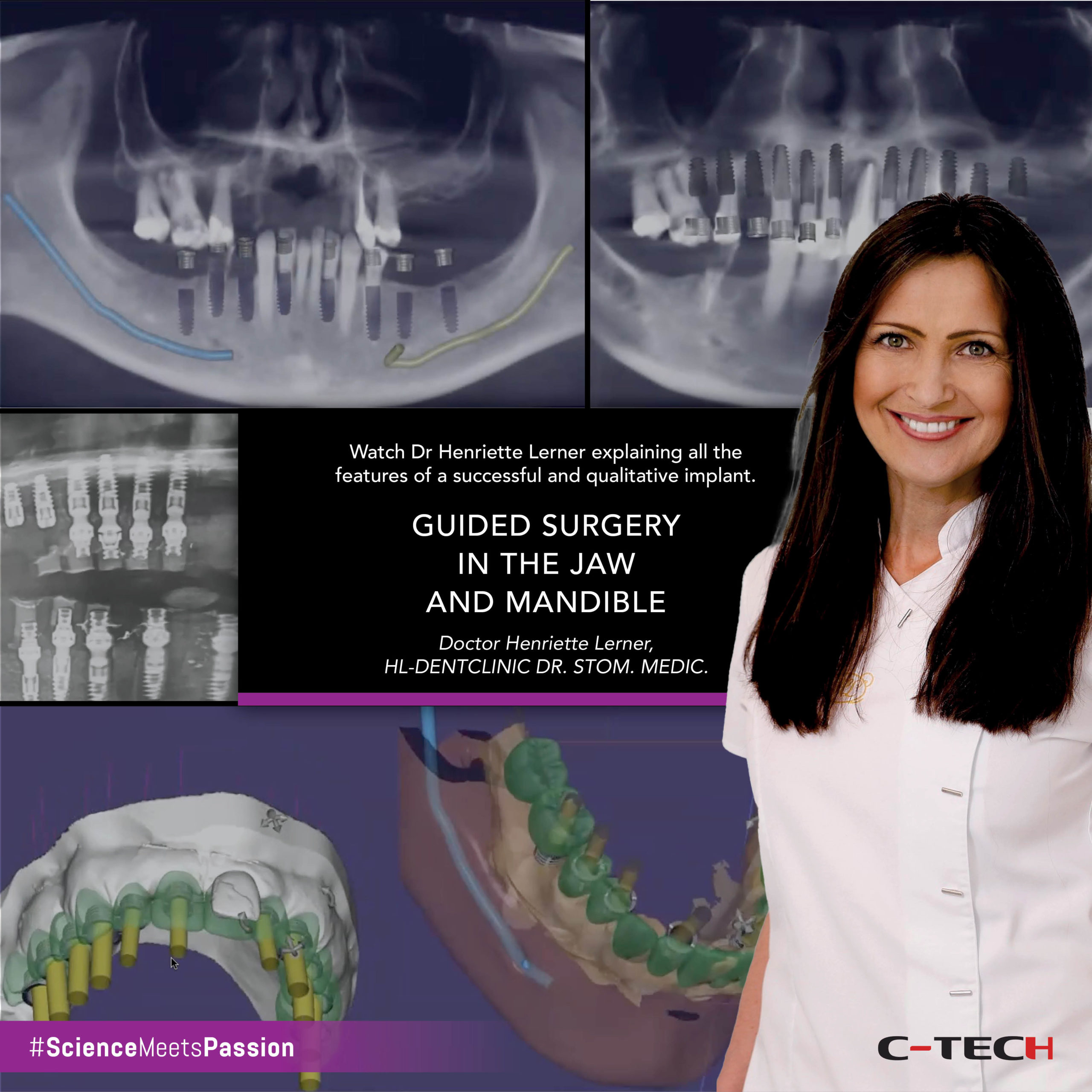
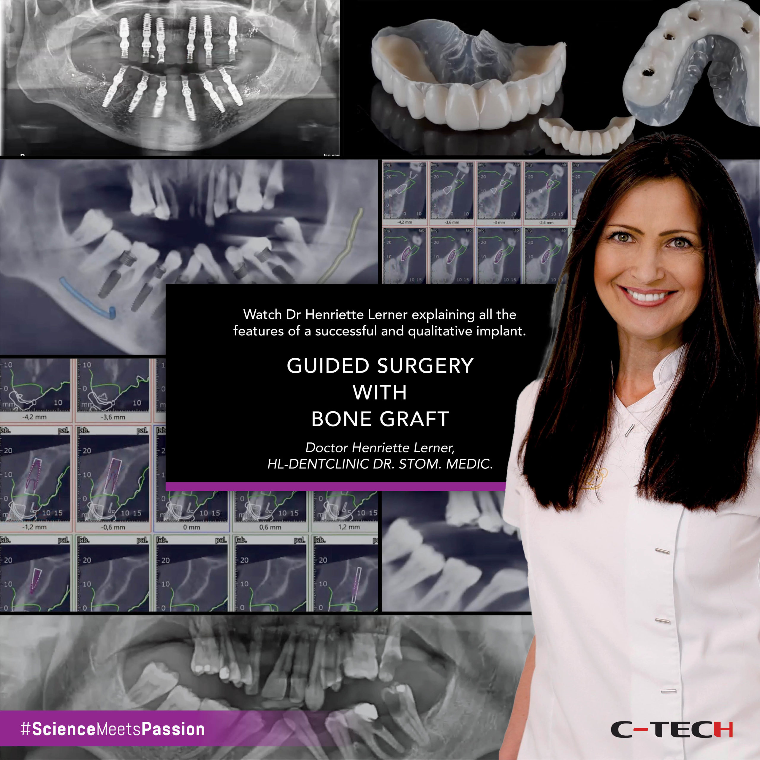
After an accurate clinical and radiological evaluation, it was decided to extract 24 and immediately place a dental implant in the extraction site with a view to immediately loading the implant with a provisional crown. Impressions were taken and an analogue wax-up was produced. To ensure the greatest accuracy, it was determined to use a digital approach to the surgery and restoration and accordingly a Cone Beam CT scan of the patient was undertaken. 3 Diemme (Serenza, Italy) Realguide planning software was chosen to plan the implant placement. The 3-dimensional imaging data captured by the CBCT scan is uploaded to the software in the form of DICOM files. The initial model from the impressions and the analogue wax up were digitally scanned and these files in .stl format were also uploaded to the 3Diemme software. The combination of these files allows for a restorative led procedure with the implant placed in an optimal position for the restoration.
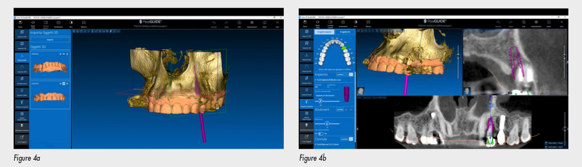
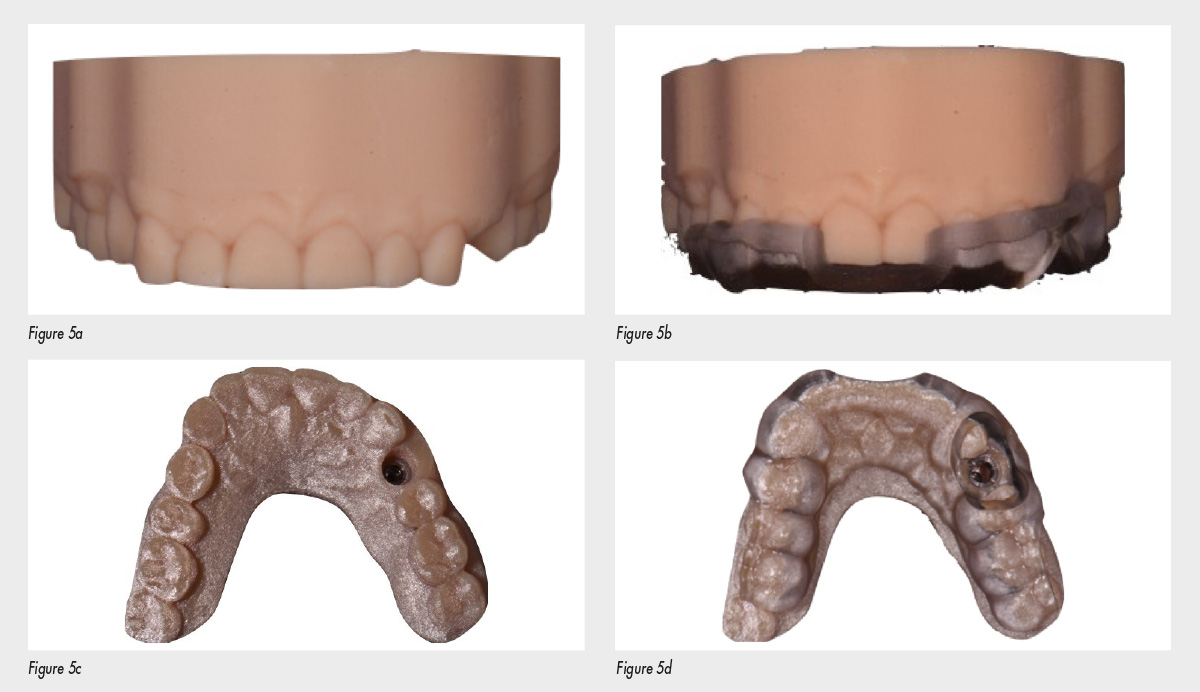
The software was also used to design the drilling template, with the appropriate guiding cylinders digitally chosen according to the implant selected Figures 4a & 4b 3Diemme planning software illustrating the combination of .stl and DICOM files to permit virtual placement of a C-Tech EL dental implant Once the virtual planning was completed in the software, an immediate visualisation of the whole process was possible including the virtual shape of the surgical guide. The files of the virtual planning and the model with the wax up were sent to 3 Diemme to produce a 3D printed model and the surgical guide.
Figures 5a & 5c 3D printed model, figures 5b & 5d model with surgical guide in place.
The C-Tech (C-Tech, Bologna, Italy) Esthetic Line (EL) implant was chosen for this case. This implant was preferred because of its modulated thread design, reducing stress from the implant to bone, thereby minimising bone resorption at the head of the implant and ensuring good primary stability at the apex. The platform switching design and sub crestal placement protocol, together with a Morse locking connection also contribute to the implant stability and reduction of marginal bone loss.
The implant placement was carried out utilising the appropriate drills according to C-Tech protocol and with a drill speed of 2000 rpm and a drilling technique that maximised irrigation of the osteotomy.
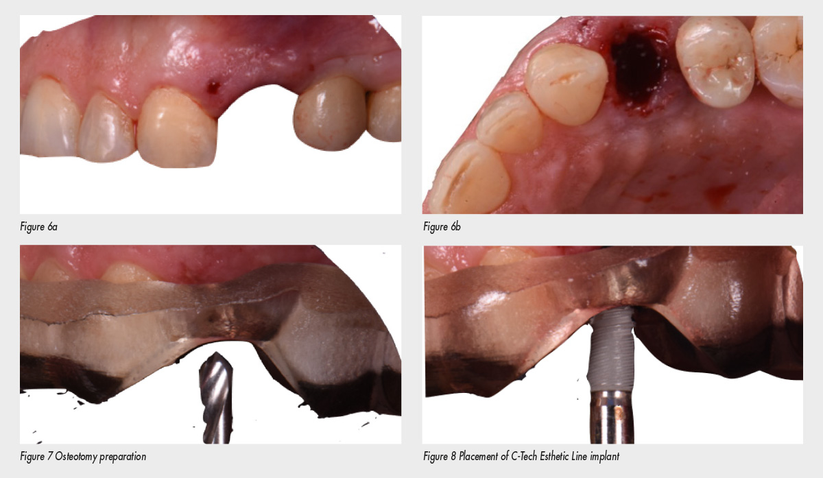
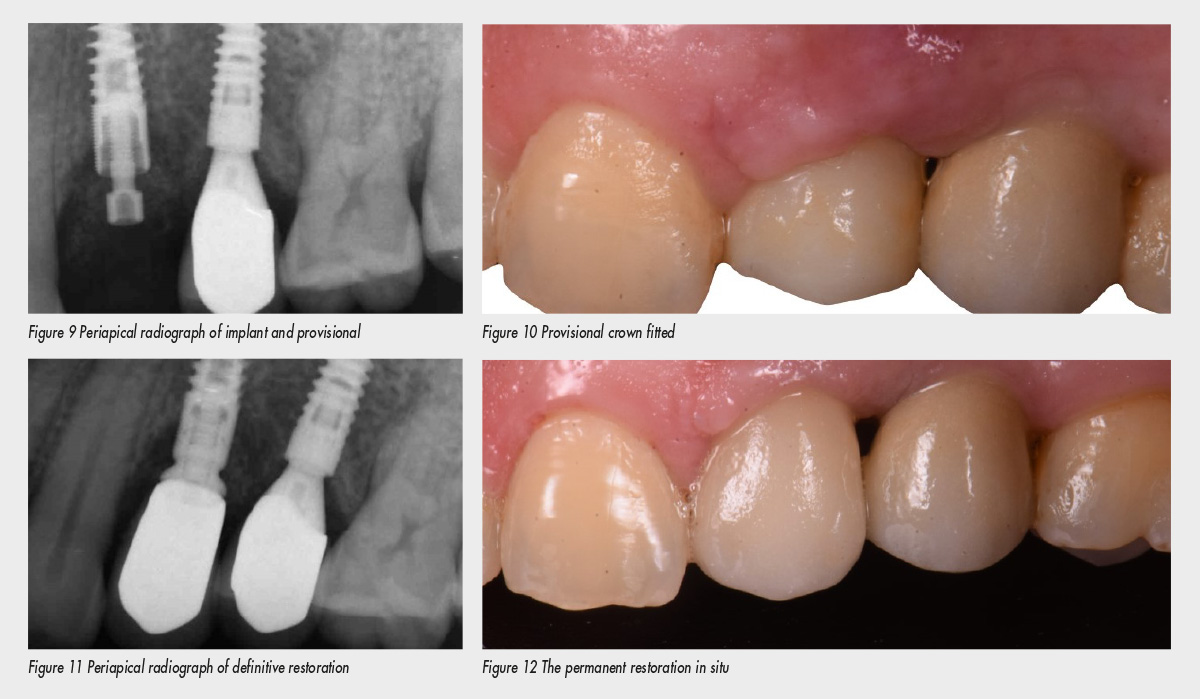
Figures 6a & 6b Root extraction prior to osteotomy preparation As planned, a 3.5mm diameter EL implant with a length of 11mm was placed. Good primary stability was achieved, satisfying the condition for immediate loading so a resin based screwretained provisional restoration was produced and was in function for three months. Before the application of this temporary crown the gap between the implant and the alveolar bone was filled with a biomaterial and a suture was applied to perfectly adapt the soft tissue to optimise the emergence profile. Three months later, the definitive crown manufactured from Zirconia/ Porcelain on a grade 5 Titanium abutment was fitted to complete the case.
Conclusion
Modern digital devices are extremely useful in improving the accuracy of treatment planning. Powerful software can combine different information basically coming from Cone Beam CT, intra oral and laboratory scanners creating a virtual environment where the clinician can easily evaluate in real time the impact of every single action on the final outcome of the therapy. At the same time, it is possible to avoid critical structures such as the ID nerve or sinus which are clearly detectable. This predictability coupled with the appropriate choice of an implant offering good primary stability can often result in immediate loading with a provisional restoration, a predictable aesthetic outcome and the faster treatment and reduced chair time contributes to increased patient satisfaction.