• Italiano
    • English
    • Deutsch
    • Español
    • Français
    • Русский
    • 中文 (中国)
    • Türkçe
    • Ελληνικά
    • Magyar
    • Српски језик
    • Lietuviškai
    • Bahasa Indonesia
    • Slovenčina

Slide Casi clinici

Henriette Lerner

Advantages of guided surgery, predictability in small spaces

Written by feRKzzCTo0 on . Posted in EL – Esthetic Line, GS – Guided Surgery, Henriette Lerner, Клинические случаи, Направленная Хирургия

by Doctor Henriette Lerner, HL-DENTCLINIC DR. STOM. MEDIC.

In this case, the patient has a genesis of the two upper lateral incisors.
Having created the space with the help of the orthodontic specialist, we first proceed with a Digital Smile Design and then with the digital planning of the two implants in comparison with the new aesthetics.

Guided surgery in the jaw and mandible

Written by feRKzzCTo0 on . Posted in EL – Esthetic Line, GS – Guided Surgery, Henriette Lerner, Клинические случаи, Направленная Хирургия

by Doctor Henriette Lerner, HL-DENTCLINIC DR. STOM. MEDIC.

In this case, it is necessary to use all the digital tools available – detection of joint movements, digital smile design and guided surgery, with the aim of having predictability and accuracy. For the future aesthetic part and function, the evaluation took place before the surgery.

Guided surgery with bone graft

Written by feRKzzCTo0 on . Posted in EL – Esthetic Line, GS – Guided Surgery, Henriette Lerner, Клинические случаи, Направленная Хирургия

by Doctor Henriette Lerner, HL-DENTCLINIC DR. STOM. MEDIC.

In this case it is shown how it is still possible to work in guided surgery and consequently perform bone grafts where necessary.
The advantage in a difficult case like this is to design the dental implants with a surgical guide, in order to maintain their correct positioning.
Finally, the aesthetic project was evaluated prior to surgery.

Webinar | EL Esthetic Line: What are the ingredients of a great implant?

Written by feRKzzCTo0 on . Posted in EL – Esthetic Line, Henriette Lerner, Клинические случаи, Традиционная Имплантология

Watch Dr Henriette Lerner explaining all the features of a successful and qualitative implant.

I saw you have breathing plant systems in the pipeline and you learn a lot of systems, and probably you already have in your mind some main features that the implants need to have.
In order to make your treatment easier and more predictable in your procedures.
You probably already know that an implant has to have a high bone implant contact surface and the designs of the implants are all made in a way that increases this surface, because this is giving you a long-term stability of the surface of the osteointegration.

Doctor Henriette Lerner | COVID-19 Guidelines

Written by feRKzzCTo0 on . Posted in Henriette Lerner, Клинические случаи, Традиционная Имплантология

How dentistry and implantology has changed in your office in phase 2?
There is no phase 1 or phase 2, there is a behavior and approach which we’ll respect in the office in the couple of years coming. Let’s start with the scheduling of the appointment of the patient: the receptionist is asking the patient about sicknesses, illnesses, cough, fever, smell alterations. The same questions she will ask 24 hours before the scheduled appointment when she calls all the patient for the next day.

Complete-arch fixed reconstruction by means of guided surgery and immediate loading: a retrospective clinical study on 12 patients with 1 year of follow-up

Written by feRKzzCTo0 on . Posted in El – Esthetic Line, Gs – Guided Surgery, Henriette Lerner, Nd – Narrow Diameter, Robert Sader, Scientific Publications, Shahram Ghanaati, Uli Hauschild, Направленная Хирургия, Традиционная Имплантология

Henriette Lerner, Uli Hauschild, Robert Sader & Shahram Ghanaati

Abstract
Guided implant surgery is considered as a safe and minimally invasive flapless procedure. However, flapless guided surgery, implant placement in post-extraction sockets and immediate loading of complete-arch fixed reconstructions without artificial gum are still not throughly evaluated. The aim of the present retrospective clinical study was to document the survival and success of complete-arch fixed reconstructions without artificial gum, obtained by means of guided surgery and immediate loading of implants placed also in fresh extraction sockets.

International Journal of Implant Dentistry – August 2017 – Исследование состояния периимплантатных тканей и стабильности периимплантатных тканей в имплантатах, вживленных с одновременной аугментацией: анализ 3-годичного ретроспективного наблюдения недавно разработанной системы имплантата на уровне кости.

Written by feRKzzCTo0 on . Posted in Century-Gtr, El – Esthetic Line, Henriette Lerner, Robert Sader, Scientific Publications, Shahram Ghanaati, Регенерация Кости, Традиционная Имплантология

International Journal of Implant Dentistry • August 2017

Исследование состояния периимплантатных тканей и стабильности периимплантатных тканей в имплантатах, вживленных с одновременной аугментацией: анализ 3-годичного ретроспективного наблюдения недавно разработанной системы имплантата на уровне кости.

Jonas Lorenz University Hospital Frankfurt · Department of oral, maxillofacial and plastic surgery, Henriette Lerner HL DENTCLINIC, Robert A. Sader Goethe-Universität Frankfurt am Main · Center of Stomatology
and Shahram Ghanaati Goethe University of Frankfurt/Main; Universitätsmedizin der Johannes Gutenberg-Universität Mainz · Department for Oral, Craniomaxillofacial and Facial Plastic Surgery; Institute of Pathology

Целью настоящего ретроспективного анализа была оценка состояния периимплантатных тканей и документирование стабильности периимплантатных тканей в имплантатах C-Tech при одновременном вживлении с аугментацией НКР (направленная костная регенерация).

47 имплантатов, которые были вживлены одновременно с аугментацией НКР (направленная костная регенерация) с синтетическим заменителем костной ткани 20 пациентам, исследовались клинически и радиологически минимум 3 года после вживления.

Последующее наблюдение показало приживаемость имплантатов 100 % и только низкие средние значения глубины зондирования (2,7 мм) и кровотечения при зондировании (30 %). Среднее значение по шкале розовой эстетики — 10,1, максимальное значение — 14.
Очевидных костных периимплантатных дефектов обнаружено не было. Среднее значение потери кости составляло 0,55 мм.

В заключение следует отметить, что имплантаты, вживленные в сочетании с процедурой НКР (направленная костная регенерация), могут достичь долгосрочного стабильного функционирования и эстетически удовлетворительных результатов при замене отсутствующих зубов в случае атрофии альвеолярного гребня.

References

1. Gurgel BC, Montenegro SC, Dantas PM, Pascoal AL, Lima KC, Calderon PD. Frequency of peri-implant diseases and associated factors. Clin Oral Implants Res. 2016; doi: 10.1111/clr.12944

2. Qian J, Wennerberg A, Albrektsson T. Reasons for marginal bone loss around oral implants. Clin Implant Dent Relat Res. 2012;14(6):792–807.

3. Berglundh T, Lindhe J, Ericsson I, Marinello C, Liljenberg B, Thomsen P. The soft tissue barrier at implants and teeth. Clin Oral Implants Res. 1991;2:81–90.

4. Berglundh T, Lindhe J, Jonsson K, Ericsson I. The topography of the vascular systems in the per iodontal and peri-implant tissues in the dog. J Clin Periodontol. 1999;21:189–93.

5. Moon I, Berglundh T, Abrahamsson I, Linder E, Lindhe J. The barrier between the keratinized mucosa and the dental implant. An experimental study in the dog. J Clin Periodontol. 1999;26:658–63.

6. Lindhe J, Berglundh T. The interface between the mucosa and the implant. Periodontol. 1998;17:47–54.

7. Masaki C, Nakamoto T, Mukaibo T, Kondo Y, Hosokawa R. Strategies for alveolar ridge reconstruction and preservation for implant therapy. J Prosthodont Res. 2015;59(4):220–8.

8. Damien CJ, Parsons JR. Bone graft and bone graft substitutes: areview of current technology and applications. J Appl Biomater.1991;2:187–208.

9. Cordaro L, Torsello F, Miuccio MT, di Torresanto VM, Eliopoulos D. Mandibular bone harvesting for alveolar reconstruction and implant placement: subjective and objective cross-sectional evaluation of donor and recipient site up to 4 y
ears. Clinical Oral Impl Res. 2011;22:1320–6.

10. Canullo L, Penarrocha-Oltra D, Soldini C, Mazzocco F, Penarrocha M, Covani U. Microbiological assessment of the implant-abutment interface in different connections: cross-sectional study after 5 years of functional loading. Clin Oral Implants Res. 2015;26(4):426–34.

11. Misch C. Implant design considerations for the posterior regions of the mouth. Implant Dent. 1999;8(4).

12. SteigengaJ,al-ShammariK,NocitiF,MischC,WangH.Dentalimplant design and its relationship to long-term implant success. Implant Dent. 2001;12(4):306–17.

13. Canullo L, Pace F, Coelho P, Sciubba E, Vozza I. The influence of platform switching on the biomechanical aspects of the implant-abutment system. A three dimensional finite element study. Med Oral Patol Oral Cir Bucal. 2011;16(6):852–6.

14. Lerner H, Lorenz J, Sader R, Ghanaati S. Two-year retrospective study of periimplant health and periimplant bone stability after immediate implant placement of a newly developed bone level implant system—a first report.
EDI Journal (European Association of Dental Implantologists, Teamwork Media); 2017; ahead of print.

15. Ghanaati S, Lorenz J, Obreja K, Choukroun J, Landes C, Sader R. Nanocrystalline hydroxyapatite-base d material already contributes to implant stability after 3 months: a clinical and radiologic 3-year follow-up investigation. In: Journal of Or al Implantology. 2014;40(1):103–9.

16. Lorenz J, Kubesch A, Korzinskas T, Barbeck M, Landes C, Sader R, et al. TRAP-positive multinucleated giant cells are foreign body giant cells rather than osteoclasts: results from a split-mouth study in humans. J Oral Implantol. 2015;41(6):e257–66.

17. Barbeck M, Udeabor S, Lorenz J, Schlee M, Grosse Holthaus M, Raetscho N, et al. High-temperature sintering of xenogeneic bone substitutes leads to increased multinucleated giant cell formation: in vivo and preliminary clinical results. J Oral Implantol. 2015;41(5):e212–22.

18. Barbeck M, Udeabor S, Lorenz J, Kubesch A, Choukroun J, Sader R, et al. Induction of multinucleated giant cells in response to small sized bovine bone substitute (Bio-Oss TM) results in an enhanced early implantation bed vascularization. Ann Maxillofac Surg. 2014;4(2):150–7.

19. Lorenz J, Barbeck M, Sader R, Russe P, Choukroun J, Kirkpatrick CJ, et al. Foreign body giant cell related encapsulation of a synthetic material three years after augmentation. J Oral Implantol. 2016;42(3):273–7.

Erfolgsfaktoren für die Behandlung mit MiniImplantaten und ihre Bedeutung für die Praxis – Praktische Implantologie und Implantatprothetik | pip 4 | 2010

Written by feRKzzCTo0 on . Posted in Henriette Lerner, Scientific Publications, Sd-Mb – Monoblock - Small Diameter, Мини-Импланты, Традиционная Имплантология

Praktische Implantologie und Implantatprothetik | pip 4 | 2010

Erfolgsfaktoren für die Behandlung mit MiniImplantaten und ihre Bedeutung für die Praxis. Prospektive Untersuchung von Patientenfällen über ein Jahr

Literatur bei den Verfassern: Henriette Lerner, Ady Palti

Der klinische Erfolg von Mini-Implantaten hängt von verschiedenen Parametern ab, die auch untereinander in Beziehung stehen. In der vorliegenden Studie wurde untersucht, ob eine Korrelation zwischen dem Durchmesser der inserierten Implantate und der Primärstabilität existiert. Dabei wurden zudem unterschiedliche, durch den jeweiligen Patientenfall vorgegebene Knochendichten berücksichtigt. Außerdem erfolgte eine Dokumentation der Osseointegration und der Tiefe etwaiger perioimplantärer Taschen über einen Zeitraum von einem Jahr post implantationem. Darüber hinaus wurden der Einfluss des Implantatdurchmessers und der Art des Implantataufbaus auf die Erfolgsrate ermittelt.

Schlussfolgerung

Aufgrund der im Rahmen der vorliegenden Untersuchung ermittelten Ergebnisse darf bei einer Verwendung von Mini-Implantaten zur Prothesenstabilisierung mit einer Erfolgsrate ähnlich wie bei klassischen Implantationen gerechnet werden. Da der Erfolg mit der Primärstabilität korreliert, lässt er sich nach deren Bestimmung mit der Drehmomentratsche direkt im Anschluss an die Insertion bereits gut abschätzen. Liegen die ermittelten Zahlen im grenzwertigen Bereich (≈ 35 Ncm), so sollte im Zweifelsfalle eine weiche Unterfütterung vorgenommen werden. Je nach individuellem Fall ist auch zu prüfen, ob zur besseren Stabilisierung ein weiteres Mini-Implantat inseriert werden kann. Hat man die Wahl, so sollte eher ein etwas größeres gewählt werden, d.h. statt des 1,8-mm-Implantats lieber das 2,1er bzw. statt des 2,4-mm-Implantats lieber das MDI Hybrid mit 2,9 mm Durchmesser.

Es versteht sich von selbst, dass eng beieinander liegende Recall-Termine angeraten sind, insbesondere um die planmäßige Osseointegration zeitnah verfolgen zu können. Sie ist keinesfalls nach sechs Monaten abgeschlossen, sondern erfährt in den darauffolgenden sechs Monaten in der Regel noch einmal eine signifikante Verbesserung.

Dr. medic.stom. Henriette Lerner

1990 Studium der Zahnmedizin (Universität für Medizin und Pharmazie „Victor Babes“ Temeschburg).
1990-1993 Oralchirurgische Weiterbildung an der Akademie für Zahnärztliche Fortbildung Karlsruhe.
1995 Training in Goldman School of Dental Implantology/Boston, Massachussets.
1998 Spezialist DGZI.
2004 Expert Implantologie der DGOI.
2006-2007 Spezialisierung “Dento-alveoläre Chirurgie” Dr. medic.stom. Henriette Lerner (Universität “Carol Davila” Bukarest).
2006 Praxis im Videnti Zentrum für Implantologie und Ästhetik, Baden Baden.
Mitglied in: DGOI; ICOI; EAO; ASA DGÄZ; DGZMK; BDO; EFOSS.
Nationale und Internationale Referententätigkeit über Ästhetik in der Implantologie, Minimal Invasive Implantology, Curriculum Implantologie, fortgeschrittene Augmentationstechniken.

Aspects of oral morphology as decision factors in mini-implant supported overdenture – Romanian Journal of Morphology and Embryology • 2010, 51(2):309–314

Written by feRKzzCTo0 on . Posted in Henriette Lerner, Scientific Publications, Sd-Mb – Monoblock - Small Diameter, Мини-Импланты, Традиционная Имплантология

Romanian Journal of Morphology and Embryology • 2010, 51(2):309–314

Aspects of oral morphology as decision factors in mini-implant supported overdenture

Elena Preoteasa, Marina MeleŞcanu-imre, Cristina Teodora Preoteasa Department of Oral Diagnosis and Ergonomics “Carol Davila” University of Medicine and Pharmacy of Bucharest, Romania, Mihaela Marin Department of Prosthodontics, Henriette Lerner Private practice, Baden Baden, Germany

Abstract

Evaluation of some morphological oral aspects perceived as decision factors in complete edentulism treatment by mini-implants overdenture. Patients, Material and Methods: An observational study was conducted on a sample of 24 patients (average age of 61 years), through clinical and imagistic methods. The variables taken into consideration were: age, gender, alveolar mucosa status, bone offer, miniimplants characteristics, insertion torque and loading type. Results: 117 mini-implants were applied. Conclusions: Mini-implant supported overdenture can be an alternative to conventional denture and conventional implant overdenture. Its advantages derives from implants’ characteristics (smaller diameter, variable length, O-ring retention system), which adapts better to the particular edentulous conditions. Insertion technique implies less surgical trauma. Choosing implants’ size, number, topography, and the loading method have a great variety, depending on anatomical feature (bone offer, mucosa and relationship with the nearby anatomical structures), functional features and patients’ wishes.

Conclusions

Mini-dental implant supported overdenture can be in complete edentulous patients a treatment alternative to both conventional dentures and conventional implant retained overdenture.

The advantages of this type of treatment derives from the characteristics of this type of implant (small diameter, variable length, O-ring retention system), which adapts better to the particular morphological conditions present in full edentulous patients. Also, the implants insertion requires less surgical trauma, this being a beneficial aspect in the context of usually poor general status.

Proceeding and achieving the treatment requires needs careful evaluation through clinical, imagistic and laboratory methods. Frequently there are identified some unfavorable conditions for implant insertion and a high degree of treatment difficulty. These issues (morphological and functional features, related to general health status, age, gender, etc.) must be linked to implants characteristics, in order to decide the particular treatment features, from surgical and prosthetic point of view. The length, diameter, number, topography, loading method of implants can present a large variety, depending on quantitative bone offer (ridge width and bone height), quality (bone density), functional features and patient’s wishes.

This type of treatment has a lower cost compared to conventional implant supported overdenture, by the lower cost of mini-implants, and also due to the use, in general, just of the panoramic radiography as imagistic method (computed tomography is an adjuvant method, but not essential in most cases). Also by eliminating some surgical intervention, we eliminate also their costs. On the other hand, the benefits related to an improved stability, better functionality and adaptation are quickly noticed by the patient and increases their level of satisfaction.

Due to the relatively simple technique, less traumatic, but with benefits that are quickly perceived, the mini-implant supported overdenture may be the elective treatment alternative for complete edentulous patients.

References

[1] PREOTEASA E, BĂNCESCU G, LONESCU E, BĂNCESCU A, DONCIU D, Epidemiologic aspects of the totally edentulous mouth. (1) General aspects, Bacteriol Virusol Parazitol Epidemiol, 2004, 49(3–4):115–120.

[2] PREOTEASA E, LONESCU E, BĂNCESCU G, BĂNCESCU A, PREOTEASA CT, Epidemiologic aspects of the edentulous mouth (II). General and local features of the totally edentulous mouth, Bacteriol Virusol Parazitol Epidemiol, 2005, 50(1–2):27–34.

[3] MELESCANU M, PREOTEASA E, Mandibular panoramic indexes predictors of skeletal osteoporosis for implant therapy, Curr Health Sci J, 2009, 35(4):291–296.

[4] FRIEDLANDER AH, The physiology, medical management and oral implications of menopause, J Am Dent Assoc, 2002, 133(1):73–81.

[5] ROBERTS WE, SIMMONS KE, GARETTO LP, DECASTRO RA, Bone physiology and metabolism in dental implantology: risk factors for osteoporosis and other metabolic bone diseases, Implant Dent, 1992, 1(1):11–21.

[6] LERNER H, Minimal invasive implantology with small diameter implants, Implant Pract, 2009, 2(1):30–35.

[7] ***, IMTEC Corporation official website, retrieved December 13, 2009, http://www.imtec.com/europe/.

[8] GRIFFITTS TM, COLLINS CP, COLLINS PC, Mini dental implants: an adjunct for retention, stability, and comfort for the edentulous patient, Oral Surg Oral Med Oral Pathol Oral Radiol Endod, 2005, 100(5):e81–e84.

[9] BALKIN BE, STEFLIK DE, NAVAL F, Mini-dental implant insertion with the auto-advance technique for ongoing applications, J Oral Implantol, 2001, 27(1):32–37.

[10] FROUM SJ, SIMON H, CHO SC, ELIAN N, ROHRER MD, TARNOW DP, Histological evaluation of bone-implant contact of immediately loaded transitional implants after 6 to 27 months, Int J Oral Maxillofac Implants, 2005, 20(1):54–60.

[11] MORNEBURG TR, PRÖSCHEL PA, Success rates of microimplants in edentulous patients with residual ridge resorption, Int J Oral Maxillofac Implants, 2008, 23(2):270–276.

[12] LABARRE EE, AHLSTROM RH, NOBLE WH, Narrow diameter implants for mandibular denture retention, J Calif Dent Assoc, 2008, 36(4):283–286.

[13] ORDOÑEZ A, EASTMOND V, Mini-dental implants – they’re here to stay, Postgraduate Dentist Caribbean, 3(1):7–12.

[14] SHATKIN TE, SHATKIN S, OPPENHEIMER AJ, Mini dental implants for the general dentist: a novel technical approach for small-diameter implant placement, Compendium, 2003, 24:26–34. st [15] MISCH CE, Dental implant prosthetics, 1 edition, Mosby, St. Louis, 2005, 130–141.

Emerging Trends in Oral Health Sciences and Dentistry – 2015 – Narrow Diameter and Mini Dental Implant Overdentures

Written by feRKzzCTo0 on . Posted in Henriette Lerner, Scientific Publications, Sd-Mb – Monoblock - Small Diameter, Мини-Импланты

Emerging Trends in Oral Health Sciences and Dentistry • 2015

Narrow Diameter and Mini Dental Implant Overdentures

https://www.intechopen.com/books/emerging-trends-in-oral-health-sciences-and-dentistry/narrow-diameter-and-mini-dental-implant-overdentures
Elena Preoteasa Department of Prosthodontics, Faculty of Dental Medicine, Carol Davila University of Medicine and Pharmacy, Bucharest, Romania, Marina Imre, Department of Prosthodontics, Faculty of Dental Medicine, Carol Davila University of Medicine and Pharmacy, Bucharest, Romania Henriette Lerner, Private Practice, Baden-Baden, Germany Ana Maria Tancu Department of Prosthodontics, Faculty of Dental Medicine, Carol Davila University of Medicine and Pharmacy, Bucharest, Romania and Cristina Teodora Preoteasa, Department of Oral Diagnosis, Ergonomics, Scientific Research Methodology, Faculty of Dental Medicine, Carol Davila University of Medicine and Pharmacy, Bucharest, Romania

Introduction

Complete dentures are most frequently a challenge for practitioners. The complexity of this disease is often associated with general health problems, but also with the physiological ageing phenomenon, that increases the treatment difficulty. Completely edentulous patients, usually elderly, often complain about the functionality of conventional dentures, especially the mandibular ones, claiming their instability, poor retention and discomfort during wear. Complete maxillary and mandibular dentures have been for over 100 years the standard treatment of complete edentulism. If complete maxillary denture wearers tolerate better the complete dentures, given the better conditions for support, retention and stability, the tolerance of mandibular prosthesis is generally lower. The relatively frequent instability of the mandibular denture, poor retention and associated discomfort were the starting point for the idea of setting the overdenture on 2 implants as first treatment alternative for the mandibular complete edentulism (according to McGill and York consensus) [2, 3, 4].

Concept of implant overdentures

Implant overdentures are inspired, as treatment concept, from the of the overdentures, the dental implants being used instead of tooth roots. Dental implants that are used for implant overdentures are made of high-strength alloy (TiAl-V), with good biocompatibility, with different designs and sizes that aim to address the prosthetic needs according to the oral particularities and clinical limitations of its execution. The first implants that were introduced in the dental practice were the ones with standard diameter, around 3.75mm. Later on, their diameter was increased and decreased (narrow), ranging between 3 and 6mm. Afterwards, the mini implants with one-piece design for implant overdentures appeared (IMTEC, later 3MESPE), with diameters of 1.8mm, 2.1mm and 2.4mm. Narrow Diameter Implant Overdenture (NDIO) represents a category of implants that combines features from conventional implants and mini implants, with diameters between 3 and 3.5mm and variable lengths (10-18mm), comprising two distinctive subgroups, namely two-piece design (e.g. Seven Narrow Line implants, MIS Implants Technologies Inc. 18-00 Fair Lawn Ave. Fair Lawn, NJ 07410, UNITED STATES, mini Sky 2, Bredent Medical GmbH & Co, Germany, Straumann implant, Straumann Group SIX: STMN, Basel Switzerland) and one-piece design (e.g. uno line, MIS implants). Two-piece narrow implants can be used as the conventional implants (with delayed loading), or as one-piece mini implants (with immediate loading protocol). In relation to anatomical, functional and prosthetic case particularities, the number of dental implants used can be reduced, similar to that of the conventional implants (e.g., two narrow implants for the mandibular overdenture). Mini Dental Implant Overdentures (MDIO) use mostly-one piece dental implants (miniSky1, Bredent, MDI 3MESPE) with diameters between 1,8mm and 3mm and variable lengths (10mm-18mm), that require one-stage surgery for implant placement, followed by prosthesis application in the same appointment, with soft material in the housing area (progressive loading) or fixation of the matrices in the denture base (immediate loading). Within the mini implants, those with a diameter between 2.7 and 3mm are classified as hybrid implants, these having sometimes a two-piece design and can be used as narrow dental implants (e.g., two narrow implants for the mandibular overdenture). The main features of the overdentures on dental implants with a diameter below the conventional one, considering their three main categories according to their diameter, are synthesized in table 1. The decision to use either a CDIO, NDIO or MDIO as treatment for complete edentulism, starts from the acknowledgment of patient’s preferences and expectations, within the limitations of the systemic and oral health-status. In systemic alterations with indications of limited surgery or that negatively affects the healing process, NDIO and MDIO are more indicated than CDIO, due to their reduced invasiveness. Oral particularities, such as the anatomical conditions (bone quality and quantity, the shape of the alveolar ridge, skeletal class), thickness and health of the oral mucosa (e.g., denture stomatitis, candidiasis), available prosthetic restorative space (especially as vertical dimension, given the necessary space for abutment, attachments and prosthesis thickness, in order to prevent its fracture) should all be considered when choosing between the implant prosthesis alternatives.

Conclusions

Stabilization of conventional denture with mini- or narrow-dental implants is beneficial especially for the elderly, considering the improvement achieved through a relatively easy surgical intervention, with moderate treatment costs. In this regard, for mandibular denture stabilization either 4 mini implants or 2 hybrid/narrow implants can be used. Treatment success is strongly related to acknowledgement of patient anatomical and functional particularities, rigorous planning and execution of prosthetic and surgical phase, as well as ensuring an adequate maintenance. Considering that edentulism is and most probably will continue to remain a frequent medical condition mostly found in the elderly, MDIO and NDIO overdentures, through their specific parameters, may replace in time complete dentures and may be the most used treatment alternative.

References

[1] WHO. Active Aging. A Policy Framework. Madrid; 2002. http://whqlibdoc.who.int/hq/2002/WHO_NMH_NPH_02.8.pdf?ua=1 (accessed 3 October 2014).

[2] Thomason JM, Kelly SA, Bendkowski A, Ellis JS. Two implant retained overdentures–a review of the literature supporting the McGill and York consensus statements Journal of Dentistry 2012;40(1) 22-34..

[3] Feine JS, Carlsson GE, Awad MA, Chehade A, Duncan WJ, Gizani S, Head T et al. The McGill Consensus Statement on Overdentures. Montreal, Quebec, Canada. May 24-25, 2002. International Journal of Prosthodont 2002;15(4) 413-4.

[4] Melescanu Imre M, Marin M, Preoteasa E, Tancu AM, Preoteasa CT.Two implant overdenture–the first alternative treatment for patients with complete edentulous mandible. Journal of Medicine and Life 2011;4(2) 207-9.

[5] Preoteasa E, Marin M, Imre M, Lerner H, Preoteasa CT. Patients’ Satisfaction With Conventional Dentures and Mini Implant Anchored Overdentures. Revista MedicoChirurgicala a Societatii de Medici si Naturisti din Iasi 2012;116(1) 310-16.

[6] Klein MO, Schiegnitz E, Al-Nawas B. Systematic review on success of narrow-diameter dental implants. The International Journal of Oral & Maxillofacial Implants 2014;29 Supplement 43-54.

[7] Diz P, Scully C, Sanz M. Dental Implants in the Medically Compromised Patient. Journal of Dentistry 2013;41(3) 195-206.

[8] Gomez-de Diego R, Mang-de la Rosa M, Romero-Pérez MJ, Cutando-Soriano A, Lopez-Valverde-Centeno A. Indications and Contraindications of Dental Implants in Medically Compromised Patients: Update. Medicina Oral Patologia Oral y Cirugia Bucal 2014;19(5):e438, -9.

[9] Preoteasa E, Murariu CM, Ionescu E, Preoteasa CT. Acrylic Resin Reinforcement With Metallic and Nonmetallic Inserts. Revista Medico-Chirurgicala a Societatii de Medici si Naturalisti din Iasi 2007; 111(2) 487-93.

[10] Lerner H. Minimal invasive implantology with small diameter implants. Implant Practice 2009, 2(1) 30-5.

[11] Preoteasa E, Meleşcanu-Imre M, Preoteasa CT, Marin M, Lerner H. Aspects of oral morphology as decision factors in mini-implant supported overdenture. Romanian Journal of Morphology and Embryology 2010;51(2) 309-14.

[12] Shatkin TE, Shatkin S, Oppenheimer AJ, et al. A simplified approach to implant dentistry with mini dental implants. Alpha Omega. 2003; 96(3) 7-15.

[13] Preoteasa E, Imre M, Preoteasa CT. A 3-Year Follow-up Study of Overdentures Retained by Mini–Dental Implants. The International Journal of Oral & Maxillofacial Implants 2014; 29(5) 1034-41.

[14] Sohrabi K, Mushantat A, Esfandiari S, Feine J. How successful are small-diameter implants? A literature review. Clinical Oral Implants Research 2012;23 (5) 515–525.

[15] Block MS1, Delgado A, Fontenot MG.The effect of diameter and length of hydroxylapatite – coated dental implants on ultimate pullout force in dog alveolar bone. Journal of Oral and Maxillofacial Surgery 1990;48(2) 174-8.

[16] Renouard F, Nisand D. Impact of implant length and diameter on survival rates. Clinical Oral Implants Research 2006;17 (2) Supplement 35-51.

[17] Singh RD, Ramashanker, Chand P. Management of atrophic mandibular ridge with mini dental implant system. National Journal of Maxillofacial Surgery 2010;1(2) 176-8.

[18] Griffitts TC, Collins CP, Collins PC. Mini dental implants: an adjunct for retention, stability, and comfort for the edentulous patient. Oral Surgery, Oral Medicine, Oral Pathology, Oral Radiology, and Endodontology 2005;100 (5) 81-4.

[19] Misch CE. Contemporary Implant Dentistry 2nd edition. St. Louis: Mosby Inc; 1999.

[20] Rossein KD. Alternative treatment plans: implant supported mandibular dentures. Inside Dentistry 2006; 2(6) 42-43.

[21] Melescanu Imre M, Preoteasa E, Tancu A, Preoteasa CT. Imaging Technique for the Complete Edentulous Patient Treated Conventionally or With Mini Implant Overdenture. Journal of Medicine and Life 2013;6(1) 86-92.

[22] Campelo LD, Camara JR. Flapless implant surgery: A 10-year clinical retro- spective analysis. International Journal Oral Maxillofacial Implants 2002;(17) 271–276.

[23] Sunitha RV, Sapthagiri E. Flapless implant surgery: A 2-year follow-up study of 40 implants. Oral Surgery, Oral Medicine, Oral Pathology and Oral Radiology 2013;116 (4) 237–243.

[24] Scherer MD, Ingel AP, Rathi N. Flapped or Flapless Surgery for Narrow-Diameter Implant Placement for Overdentures: Advantages, Disadvantages, Indications, and Clinical Rationale. The International Journal of Periodontics & Restorative Dentistry 2014;34(3) Supplement 89-95.

[25] Christensen GJ.The ‘mini’-implant has arrived. The Journal of the American Dental Association 2006;137(3) 387-90.

[26] Preoteasa E, Iosif L, Amza O, Preoteasa CT, Dumitrascu C. Thermography, an Imagistic Method in Investigation of the Oral Mucosa Status in Complete Denture Wearers. Journal of Optoelectronics and Advanced Materials 2010;12(11) 2333–4.

[27] Awad MA, Lund JP, Dufresne E, Feine JS. Comparing the efficacy of mandibular implant-retained overdentures and conventional dentures among middle-aged edentulous patients: satisfaction and functional assessment. The International Journal of Prosthodontics 2003;16, 117–22.

[28] Flanagan D, Mascolo A. The Mini Dental Implant in Fixed and Removable Prosthetics: A Review. Journal of Oral Implantology 2011;37 (1) 123-132

[29] Bulard RA. Mini implants. Part I. A solution for loose dentures. The Oklahoma Dental Association Journal. 2002;93.42-46.

[30] Dantas Ide S, Souza MB, Morais MH, Carreiro Ada F, Barbosa GA. Success and survival rates of mandibular overdentures supported by two or four implants: a systematic review, Brazilian Oral Research 2014;28(1) 74-80.

[31] Bergendal T, Engquist B. Implant-supported overdentures: a longitudinal prospective study. The International Journal of Oral & Maxillofacial Implants 1998;13 (2) 253–62.

[32] Klein MO, Schiegnitz E, Al-Nawas B. Systematic review on success of narrow-diameter dental implants. The International Journal of Oral & Maxillofacial Implants. 20

Implant Practice – February 2009 Volume 2 Number 1 – Minimal invasive implantology with small diameter implants

Written by feRKzzCTo0 on . Posted in Henriette Lerner, Scientific Publications, Sd-Mb – Monoblock - Small Diameter, Мини-Импланты

Implant Practice • February 2009 Volume 2 Number 1

Minimal invasive implantology with small diameter implants

Henriette Lerner

What are mini implants?

The highest target in our profession is the fulfilment of patient wishes. The greatest wish of our patient is always the fast, painless replacement of their missing teeth or stabilisation of the prosthesis. A fast, stable and esthetic reconstruction of the patient’s dento-facial system is the main goal of every dentist.

At the time, immediate loading was not an issue, the mini implants were used for the stabilisation of a provisional construction for the time necessary for the osseointegration of the conventional implants. Those mini implants were 1.8mm to 3.3mm in diameter. This implant was also developed with a small ball on the top of it that could be incorporated as a snap for a denture, or to secure a temporary bridge. To uncover the standard implants, it was found that around 50% of the mini implants had actually integrated or bonded to the bone. In order to increase the rate of success, the implant design was improved in to follow the rules of osseointegration and the insertion protocol was changed to give the implants the primary stability necessary for immediate occlusal loading.

Indication for insertion

The general indication for the placement of narrow diameter implants,ridges which, through resorption, become inadequate for placement of standard diameter implants: buco-lingually <5mm, mesiodistally <5mm or both.

The absolute indication will be completed in these cases where the patient does not want an augmentative bone reconstruction or is not indicated from the medical point of view.
a) Edentulous arches The indication for the lower jaw is a 1.8-2.1mm diameter implant. For the insertion of an implant with a diameter of 1.8-2.1mm we need 10mm bone height and 3mm bone width. For the upper jaw the recommended implant is 2.4mm diameter. 4mm bone width and 10mm bone height will be needed.
b) Single tooth restoration The second indication is the insertion of a mini implant in a small gap, in order to replace a front tooth or a premolar, in situations where the standard diameter implant is contraindicated or not possible. A small gap of mesio distal dimension of 5 mm can be replaced with an implant of 2.4mm and a crown if the esthetical considerations will be adequate.

References

[1] Roland Glauser, Peter Schüpbach, Jan Gottlow, Christoph HF Hämmerle. Peri-implant Soft Tissue Barrier at Experimental One-Piece Mini-implants with Different Surface Topography in Humans: A Light-Microscopic Overview and Histometric Analysis. Clinical Implant dentistry and Related Research 7: issue s1

[2] Misch CE (1990). Density of bone: effect on treatment plans,surgical approach, healing and progressive bone loading. Int J Oral Implant 6:23-31

[3] Todd Shatkin, Samuel Schatkin, Benjmin D Openheimer, Adam J Openheimer (2007). Mini Dental Implants for Long-Term Fixed and Removable Prosthetics: A Retrospective Analysis of 2514 Implants Placed Over a FiveYear Period. Compendium 28 (2): 36-41

[4] Zeev Ormianer, Arun K Garg, Ady Palti (2006). Immediate Loading of Implant Overdentures Using Modified Loading Protocol. Implant dentistry 15: 1

[5] Mi-Ra Ahn, Kyung-Mi An, Jung-Hwan Choi, Dong-Seok Sohn (2004). Immediate Loading With Mini Dental Implants in the Fully Edentulous Mandible. Implant Dentistry 13:4

[6] Carl E Misch, Hom-Lay Wang, Craig M Misch, Mohamed Sharawy, Jack Lemons, Kenneth WM Judy (2004). Rationale for the application of immediate load in Implant dentistry. Implant Dentistry 13:3

[7] Sahin S, Cehreli MC, Yalcin E (2002). The influence of functional forces on the biomechanical of implants-supported prostheses – a review. J Dent 30:271-282

[8] Vigolo P, Givani A (2000). Clinical evaluation of single tooth mini-implant restaurations: a five year retrospective study , Journal of Orosthetic Dentistry July, 84(1):50-4

[9] Burto E Balkin, David E Stefik, Francie Naval (2001). Mini–dental implant insertion with the auto advance technique for onoing applications. Journal of Oral Implant 27: 1

[10] Carl E Misch, Jon B Suzuki, Francine Misch-Dietsh, artha W Bidez (2005). A Positive Correlation Between Occlusal Trauma and Peri-implant Bone Loss: Literature Support. Implant Dentistry 14: 2

[11] Cornelis N Scheffler, H De Clerck, J Tulloch, C Behets. Systematic review of the experimental use of temporary skeletal anchorage devices in orthodontics. American Journal of Orthodontics and Dentofacial Orthopedics. 131 (4): S52-S58 M

[12] Misch, Carl E; Hahn, Jack; Judy, Kenneth W; Lemons, Jack E; Linkow, Leonard I; Lozada, Jamie L; Mills, Edward; Misch, Craig M; Salama, Henry; Sharawy, Mohamed; Testori, Tiziano; Wang, Hom-Lay DOI: 10.1563/0722.1. Workshop Guidelines on Immediate Loading in Implant Dentistry. Journal of Oral Implantology Issn: 1548-1336. 30 (5): 283288

[13] Sendax VI (1995). Mini implants strategy offers a broad range of uses. Dent Today 14(1): 227–232

[14] Mishal M, De Souza, Sabita M. Ram, Kartik Bhanushali. Dept of Prosthodontics, Pad Dr. D.Y. Patil Dental College and Hospital, Nerul Management of atrophic mandibular ridges with Mini Dental Implant system – A case report

[15] Ron A Bulard (2003). Mini Dental Implants: Enhancing Patient Satisfaction and Practice income. Dentistry Today. 10: 7

[16] Trevor McClain Griffitts, Chad Patrick Collins, Patrick Charles Collins, Spokane Wash. Mini dental implants: An adjunt for retention, stability, and comfort for the edentulous patient. Oral Surg Oral Med Oral Pathol Radiol Endod 100:E81-4

[17] Ziv Mazor, Marius Steigmann, Roy Leshem, Micahel Peleg (2004). Miniimplants to Reconstruct Missing Teeth in Severe Ridge deficiency and small Interdental Space: a 5 –year case series, Implant dentistry 13: 4

[18] Campelo LD, Camara JR (2002). Flapless Implant surgery: a 10-year clinical retrospective analysis, Jomi Mar- April; 17(2):271-6

[19] Chiapasco M, Gatti C, Rossi E, Haefliger W, Markwalder TH (1997). Implant-retained mandibular overdenture with immediate loading. A retrospective multicenter study on 226 consecutive cases. Clin Oral Implants Res 8:48-57

[20] Sang-Choon Cho, Stuart Froum,Chih –Han TAi, Young Sung Cho, Nicolas Elian, Dennis Tarnow. Immediate loading of narow diameter implants with overdentures in severely atrophic mandibles. Pract Proced Aesthet Dent Apr; 19 (3):167-74

[21] Misch CE (1993). Patient force factors. In: CE Misch, ed. Contemporary Implant Dentistry. St. Louis, MO: CV Mosby

[22] Adell R, Lekholm U, Rockler B, Branemark PL (1981). A 15-year study of osseointegrated implants in the treatment of the edentulous jaw. Int J Oral Surg 10(6):387–416

[23] Branemark PL (1983). Osseointegration and its experimental background. J Prosthet Dent 50(3):399–410

[24] Dessem D, Taylor A (1989). Reflex effects of periodontal mechanoreceptors on trigeminal motoneurons. In Van Steenberbghe D, DeLaat A (eds): Electromyography of Jaw Reflexes in Man. Leuven, Belgium: Leuven University Press 177-196

EDI Journal – December 2016 – Первый отчет по двухлетнему ретроспективному исследованию с недавно разработанной системой имплантации на уровне кости Периимплантатное здоровье и периимплантатная стабильность кости после немедленного вживления имплантата Доктор стоматологии

Written by feRKzzCTo0 on . Posted in El – Esthetic Line, Henriette Lerner, Robert Sader, Scientific Publications, Shahram Ghanaati, Традиционная Имплантология

International Journal of Implant Dentistry • December 2016

Первый отчет по двухлетнему ретроспективному исследованию с недавно разработанной системой имплантации на уровне кости Периимплантатное здоровье и периимплантатная стабильность кости после немедленного вживления имплантата Доктор стоматологии

DR-Medic Stom. Henriette Lerner HL DENTCLINIC, Dr. Jonas Lorenz University Hospital Frankfurt · Department of oral, maxillofacial and plastic surgery, Professor Robert A. Sader Goethe-Universität Frankfurt am Main · Center of Stomatology and Dr. Shahram Ghanaati Goethe University of Frankfurt/Main; Universitätsmedizin der Johannes Gutenberg-Universität Mainz · Department for Oral, Craniomaxillofacial and Facial Plastic Surgery; Institute of Pathology

Стоматологические имплантаты стали надежным и прогнозируемым средством для замены отсутствующих зубов и сохранения зубного ряда у пациентов с адентией. Они могут восстановить здоровье ротовой полости, ее форму, функцию, жевание, артикуляцию и эстетику стоматологической системы — многолетняя успешность имплантации составляет более 90 % имплантатов у пациентов с полной [1,2] или частичной [3–6] адентией. Вариации в успешности имплантации зависят от техники проведения операции, протокола нагрузки, локализации имплантата и качества кости, например, о более низкой успешности имплантации сообщается при верхнечелюстной имплантации, в отличие от нижнечелюстной [7,8].

За последние десятилетия исследования стоматологических имплантатов привели к значительным изменениям хирургических и простетических протоколов.
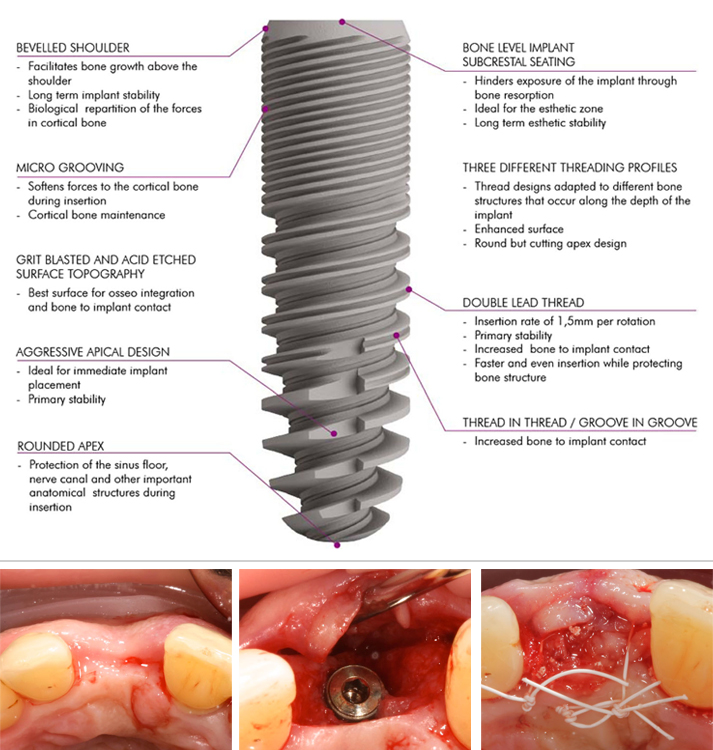
Что касается поверхности имплантатов, существует единогласное мнение о превосходстве поверхностей с шероховатостью/микротекстурой.

Другие способы улучшения поверхности имплантата включают конструкцию резьбы, длину имплантата и ширину имплантата. Конструкция имплантата должна обладать свойствами, которые наилучшим образом трансформируют натяжение и сдвигающее усилие во время жевания и минимизируют нежелательные составляющие силы.
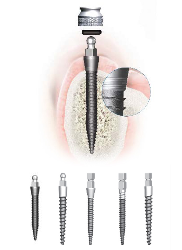
Кроме того, для долгосрочной стабильности периимплантатной костной ткани, а также эстетически и функционально удовлетворительного стоматологического имплантата необходима стабильность соединения имплантата с абатментом, чтобы предотвратить трещины имплантата и ослабление винтов и сохранить стабильность периимплантатного уровня кости.
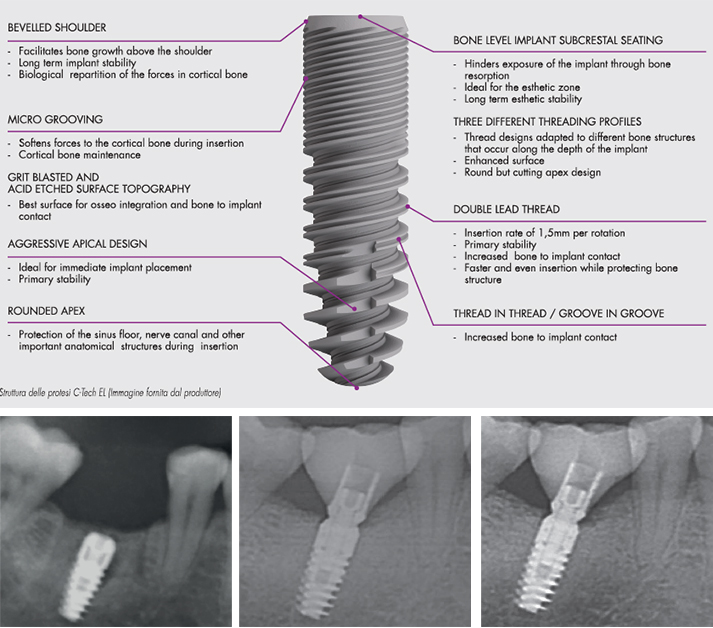
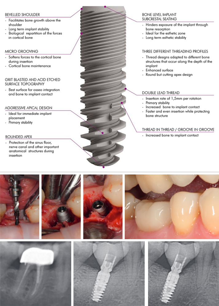
При конструкции, состоящей из двух частей, нельзя избежать расстояния или микрозазора между имплантатом и абатментом. Тем не менее меньший микрозазор иногда может образовываться в конструкциях с соединением платформ различных диаметров и в коническом соединении конусом Морзе, которые используются для передачи взаимодействия микрозазора и оси имплантата, а также для снижения микродвижений. Это может уменьшить поступательные движения жидкости десневой борозды и, следовательно, потерю альвеолярного гребня, даже если имплантаты установлены под альвеолярный гребень (ниже уровня костного гребня).

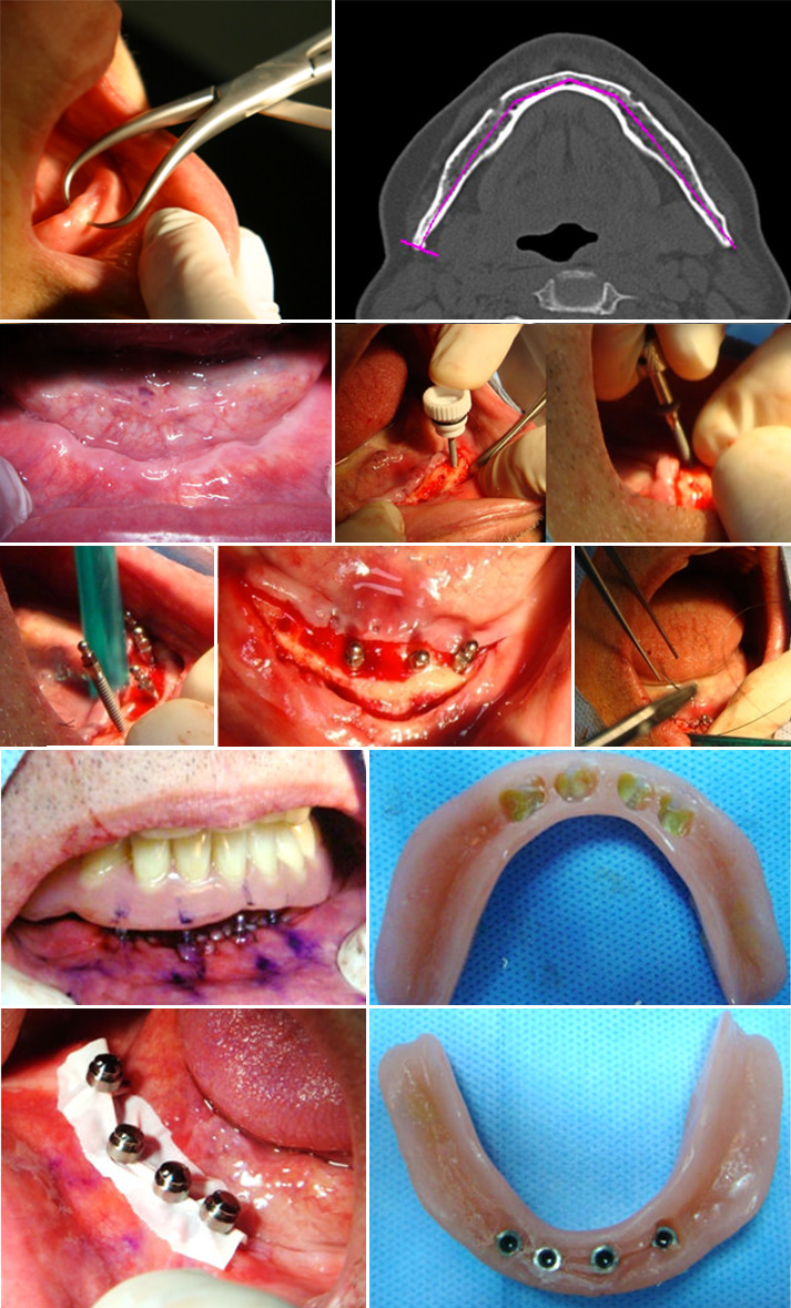
Цель этого ретроспективного исследования — описать клинические и радиологические результаты новой имплантационной системы с пескоструйно обработанной и протравленной кислотой топографией поверхности и коническим соединением Морзе имплантата и абатмента. Имплантаты были установлены в свежие и неповрежденные постэкстракционные лунки зубов верхней и нижней челюстей, которые не подлежали лечению, и наблюдались клинически и радиологически после среднего времени нагрузки на протяжении двух лет. Особое внимание было уделено поддержанию периимплантатного здоровья и стабильности периимплантатного уровня кости.


References

1. Albrektsson T, Dahl E, Enbom L, Engevall S, Engquist B, et al. (1988) Osseointegrated oral implants. A Swedish multicenter study of 8139 consecutively inserted nobelpharma implants. J Periodontol 59(5):287-296.

2. Spiekermann H, Jansen VK, Richter EJ (1995) A 10-year follow-up study of IMZ and TPS implants in the edentulous mandible using bar-retained overdentures. Int J Oral
Maxillofac Implants 10(2):231-243.

3. Nevins M, Langer B (1993) The successful application of osseointegrated implants to the posterior jaw: a long-term retrospective study. Int J Oral Maxillofac Implants 8(4):428-432.

4. Henry PJ, Laney WR, Jemt T, Harris D, Krogh PH, et al. (1996) Osseointegrated implants for single-tooth replacement: a prospective 5-year multicenter study. Int J Oral Maxillofac Implants 11(4):450-455.

5. Schmitt A, Zarb GA (1993) The longitudinal clinical effectiveness of osseointegrated dental implants for single-tooth replacement. Int J Prosthodont 6(2):197-202.

6. Fugazzotto PA, Gulbransen HJ, Wheeler SL, Lindsay JA (1993) The use of IMZ osseointegrated implants in partially and completely edentulous patients: success and failure rates of 2,023 implant cylinders up to 60+ months in function. Int J Oral Maxillofac Implants 8(6):617-621.

7. Misch CE (1990) Density of bone: effect on treatment plans, surgical approach, healing, and progressive boen loading. Int J Oral Implantol 6(2):23-31.

8. Albrektsson T, Lekholm U (1989) Osseointegration: current state of the art. Dent Clin North Am 33 (4):537-554.

9. Adell R, Lekholm U, Brånemark PI (1985) Surgical procedures. In: Brånemark PI, Zarb G, Albrektsson T (Eds.), Tissue Integrated Prostheses: Surgical Procedures, Quintessence Publishing Co, Chicago, USA, pp. 223-225.

10. Kohal RJ, LaRosa M, Patrick D, Hürzeler MB, Caffesse RG (1999) Clinical and histologic evaluation of submerged and nonsubmerged hydroxyapatite-coated im-plants: a preliminary study in dogs. Int J Oral Maxillofac Implants 14(6):824-834.

11. Evian CI, Kessler L, Axler J (1997) One-stage surgery with a nonsubmerged implant system. Compend Contin Educ Dent 18(11):1091-1094,1096-1098.

12. Ericsson I, Nilner K, Klinge B, Glantz PO (1996) Radio-graphical and histological characteristics of submerged and nonsubmerged titanium implants. An experimental study in the Labrador dog. Clin Oral Implants Res 7(1):20-26.

13. Chrcanovic BR, Albrektsson T, Wennerberg A (2015) Dental implants inserted in fresh extraction sockets versus healed sites: a systematic review and meta-analysis. J Dent 43(1):16-41.

14. Weiss CM, Weiss A, Rosenlicht J (2001) Root form implants. Treatment of total mandibular edentulism diagnosed for an overdenture. In: Weiss CM, Weiss A, (Eds.), Principles and Practice of Implant Dentistry, Mosby, St Louis, USA, pp. 147-168.

15. Fickl S, Zuhr O, Wachtel H, Stappert CF, Stein JM, et al. (2008) Dimensional changes of the alveolar ridge contour after different socket preservation techniques. J Clin Peri-odontol 35(10):906-913.

16. Vignoletti F, Matesanz P, Rodrigo D, Figuero E, Martin C, et al. (2012) Surgical protocols for ridge preservation after tooth extraction. A systematic review. Clin Oral Implants Res 23(5):22-38.

17. Lekovic V, Camargo PM, Klokkevold PR, Weinlaender M, Kenney EB, et al. (1998) Preservation of alveolar bone in extraction sockets using bioabsorbable membranes. J Periodontol 69(9):1044-1049.

18. Becker W, Becker B, Polizzi G, Bergstrom C (1994) Autogenous bone grafting of defects adjacent to implants placed into immediate extraction sockets in patients: a prospective study. Int J Oral Maxillofac Implants 9(4):389-396.

19. Artzi Z, Tal H, Dayan D (2001) Porous bovine bone mineral in healing of human extraction sockets: 2. Histo-chemical observations at 9 months. J Periodontol 72(2):152-159.

20. Iasella JM, Greenwell H, Miller RL, Hill M, Drisko C (2003) Ridge preservation with freeze-dried bone allograft and a collagen membrane compared to extraction alone for implant site development: a clinical and histologic study in humans. J Periodontol 74(7):990-999.

21. Araújo MG, Lindhe J (2005) Dimensional ridge alterations following tooth extraction. An experimental study in the dog. J Clin Periodontol 32(2):212-218.

22. Evans CD, Chen ST (2008) Esthetic outcomes of immediate implant placements. Clin Oral Implants Res 19(1):73-80.

23. Chen ST, Darby IB, Reynolds EC, Clement JG (2009) immediate implant placement postextraction without flap elevation. J Periodontol 80(1):163-172.

24. Lang NP, Pun L, Lau KY, Li KY, Wong MC (2012) A systematic review on survival and success rates of implants placed immediately into fresh extraction sockets after at least 1 year. Clin Oral Implants Res 23(5):39-66.

25. Pagni G, Pellegrini G, Giannobile WV, Rasperini G (2012) Postextraction alveolar ridge preservation: biological basis and treatments. Int J Dent 2012:151030.

26. Tavarez RR, Calixto AM, Maia Filho EM, Bandeca MC, Firoozmand LM, et al. (2014) Atraumatic extraction, implant placement and immediate provisionalization. J Contemp Dent Pract 15(4):513-517.

27. Misch CE (1999) Implant design considerations for the posterior regions of the mouth. Implant Dent 8(4):376-386.

28. Binon PP (2000) Implants and components: entering the new millennium. Int J Oral Maxillofac Implants 15(1):76-94.

29. Trisi P, Rao W, Rebaudi A (1999) A histometric comparison of smooth and rough titanium implants in human low-density jawbone. Int J Oral Maxillofac Implants 14(5):689-698.

30. Steigenga JT, Shammari KF, Nociti FH, Misch CE, Wang HL (2003) Dental implant design and its relationship to long-term implant success. Implant Dent 12(4):306-317.

31. Niznick G (2000) Achieving Osseointegration in soft bone: The search for improved results. Oral Health 90:27-32.

32. O’Sullivan D, Sennerby L, Meredith N (2000) Measurements comparing the initial stability of five designs of dental implants: a human cadaver study. Clin Implant Dent Relat Res 2(2):85-92.

33. Sykaras N, Iacopino AM, Marker VA, Triplett RG, Woody RD (2000) Implant materials, designs, and surface topographies: their effect on osseointegration. A literature review. Int J Oral Maxillofac Implants 15(5):675-690.

34. Canullo L, Penarrocha OD, Soldini C, Mazzocco F, Penarrocha M (2015) Microbiological assessment of the implant-abutment interface in different connections: cross-sectional study after 5 years of functional loading. Clin Oral Implants Res 26(4):426-434.

35. Pozzi A, Tallarico M, Moy PK (2014) Three-year post-loading results of a randomised, con-trolled, split-mouth trial comparing implants with different prosthetic interfaces and design in partially posterior edentulous mandibles. Eur J Oral Implantol 7(1):47-61.

36. Ghanaati S, Lorenz J, Obreja K, Choukroun J, Landes C, et al. (2014) Nanocrystalline hydroxyl apatite-based material already contributes to implant stability after 3 months: a clinical and radiologic 3-year followup investigation. J Oral Implantol 40(1):103-109.

37. Ghanaati S, Barbeck M, Lorenz J, Stuebinger S, Seitz O (2013) Synthetic bone substitute material comparable with xeno-geneic material for bone tissue regeneration in oral cancer patients: First and preliminary histological, histomorpho-metrical and clinical results. Ann Maxillofac Surg 3(2):126-138.

38. Sailer I, Zembic A, Jung RE, Siegenthaler D, Holderegger C (2009) Randomized controlled clinical trial of customized zirconia and titanium implant abutments for canine and posterior single-tooth implant reconstructions: preliminary results at 1 year of function. Clinical Oral Implants Research 20(3):219-225.

39. Brägger U, Bürgin WB, Hämmerle CH, Lang NP (1997) Associations between clinical parameters assessed around implants and teeth. Clinical Oral Implants Research 8(5):412-421.

40. Berglundh T, Lindhe J, Ericsson I, Marinello CP, Liljenberg B, et al. (1991) The soft tissue barrier at implants and teeth. Clinical Oral Implants Research 2(2):81-90.

41. Berglundh T, Lindhe J, Jonsson K, Ericsson I (1994) The topography of the vascular systems in the periodontal and peri-implant tissues in the dog. Journal of Clinical Periodontology 21(3):189-193.

42. Moon IS, Berglundh T, Abrahamsson I, Linder E, Lindhe J (1999) The barrier between the keratinized mucosa and the dental implant. An experimental study in the dog. Journal of Clinical Periodontology 26(10):658-663.

43. Lindhe J, Berglundh T (1998) The interface between the mucosa and the implant. Periodontology 2000 17: 47-54.

44. Rieder D, Eggert J, Krafft T, Weber HP, Wichmann MG (2014) Impact of placement and restoration timing on single-implant esthetic outcome-a randomized clinical trial. Clin Oral Implants Res 27(2):e80-e86.

45. Romanos GE, Aydin E, Locher K, Nentwig GH (2014): Immediate vs. delayed loading in the posterior mandible: a split-mouth study with up to 15 years of follow-up. Clin Oral Implants Res 27(2):e74-e79.

46. Wagenberg B, Froum SJ (2014) Long-Term Bone Stability around 312 Rough-Surfaced Immediately Placed Implants with 2-12-Year Follow-Up. Clin Implant Dent Relat Res 17(4):658-666.

47. Javed F, Ahmed HB, Crespi R, Romanos GE (2013) Role of primary stability for successful osseointegration of dental implants: Factors of influence and evaluation. Interv Med Appl Sci 5(4):162-167.

48. Calvo GJL, Gomez MG, Aguilar SA, Mate SVJE, Abboud M (2014) Bone remodeling at implants with different configurations and placed immediately at different depth into extraction sockets. Experimental study in dogs. Clin Oral Implants Res 26(5):507-515.

49. Javed F, Almas K, Crespi R, Romanos GE (2011) Implant surface morphology and primary stability: is there a connection? Implant Dent 20(1):40-46.

50. Berberi A, Tehini G, Rifai K, Bou NEF, El ZN (2014) In vitro evaluation of leakage at implant-abutment connection of three implant systems having the same prosthetic interface using rhodamine B. Int J Dent 2014:351263.

Dentista Moderno, Italia – October 2016 – Контрольное обследование состояния периимплантных тканей и устойчивости костной ткани по истечении двух лет в ряде случаев с одномоментной установкой имплантационной системы на костной ткани недавней остеопластики

Written by feRKzzCTo0 on . Posted in El – Esthetic Line, Henriette Lerner, Robert Sader, Scientific Publications, Shahram Ghanaati, Традиционная Имплантология

Dentista Moderno, Italia • October 2016

Контрольное обследование состояния периимплантных тканей и устойчивости костной ткани по истечении двух лет в ряде случаев с одномоментной установкой имплантационной системы на костной ткани недавней остеопластики.Первый доклад

Jonas Lorenz University Hospital Frankfurt · Department of oral, maxillofacial and plastic surgery, Henriette Lerner HL DENTCLINIC, Robert A. Sader Goethe-Universität Frankfurt am Main · Center of Stomatology
and Shahram Ghanaati Goethe University of Frankfurt/Main; Universitätsmedizin der Johannes Gutenberg-Universität Mainz · Department for Oral, Craniomaxillofacial and Facial Plastic Surgery; Institute of Pathology

Введение
Зубные имплантаты представляют собой надежный и предсказуемый в ортопедической стоматологии метод для замены и протезирования зубов в случае адентии. Таким образом, здоровье ротовой полости, форма, функция, акт жевания, челюстное сочленение и эстетика стоматогнатического аппарата могут быть восстановлены с вероятностью долгосрочного успеха более 90% в случаях установки зубных имплантов у пациентов с полной или частичной адентией.
Для успешной долгосрочной имплантации необходимо соблюдать некоторые специфические требования технического и конструкционного характера. Устойчивость соединения имплантата с зубной культей является еще одним существенным фактором для долгосрочной стабильности периимплантной костной ткани и для обеспечения эстетического эффекта и функциональности зубного импланта. Возникновение пространства или микроразрыва между имплантом и зубной культей неизбежно, но несмотря на это можно достигнуть наименьшего микроразрыва, если имплантационный дизайн предусматривает конусное соединение при помощи конуса Морзе и принцип platform switching (соединение платформ различного диаметра), которые перемещают микроразрыв перед имплантационной осью и ограничивают микродвижение. Поэтому нагнетание зубного ликвора и последующая потеря гребневой костной ткани могут быть уменьшены, даже в случае установки подгребневого импланта.

Этот ряд случаев представлен с целью впервые описать клинические и радиологические результаты по истечении двух лет с момента одномоментной установки 50 имплантов посредством новой имплантационной системы с пескоструйной и кислотной поверхностной обработкой и с конусным соединением Морзе.

На протяжении в среднем двухгодичного периода наблюдения у ни одного импланта не были выявлены дефекты, острые воспаления или периимплантит. Во всех имплантах выявлено достаточное количество периимплантной кератинизированной мягкой ткани, низкий процент глубины зондирования (в среднем 2,25 мм) и индекс кровоточивости 34%. После двухлетней нагрузки уровень периимплантной костной ткани стабилизировался со средней потерей костной ткани в объеме 0,83 мм.

В течение среднего двухгодичного периода наблюдения рассматриваемый имплант с одномоментной установкой на уровне костной ткани, с шероховатой поверхностью и конусным соединением показал здоровое состояние периимплантных мягких и твердых тканей. Исследуемые параметры соответствуют результатам подобных исследований, приведенных в зарубежной литературе, или превосходят их.


References

1. Albrektsson, I; Dahl, E.; Enbom, L; Engevall, S.; Engquist, B.; Eriksson, A. R. et al. (1988): Osseointegrated oral implants. A Swedish multicenter study of 8139 consecutively inserted Nobelpharma implants. In: J. Periodontal 59 (5):287-296.

2. Spiekermann, H.; Jansen, V. K.; Richter, E. J. (1995): A 10-year follow-up study of IMZ and TPS implants in the edentulous mandible using bar-retained overdentures. In: Int J Oral Maxillofac Implants 10 (2):231 -243.

3. Nevins, M.; Longer, B. (1993): The successful application of osseointegrated implants to the posterior jaw: a long-term retrospective study. In: Int J Oral Maxillofac Implants 8 (4):428-432.

4. Henry, P. J.; Laney, W. R.; Jemt, T.; Harris, D.; Krogh, P. H.; Polizzi, G. et al. (1996): Osseointegrated implants for single-tooth replacement: a prospective 5-year multicenter study. In: Int J Oral Maxillofac Implants 11 (4):450-455.

5. Schmitt, A.; Zarb, G. A. (1993): The longitudinal clinical effectiveness of osseointegrated dental implants for single-tooth replacement. In: Int J Prosthodont 6 (2):197-202.

6. Fugazzotto, P. A.; Gulbransen, H. J.; Wheeler S. L.; Lindsay, J. A. (1993): The use of IMZ osseointegrated implants in partially and completely edentulous patients: success and failure rates of 2,023 implant cylinders up to 60+ months in function. In: Int J Oral Maxillofac Implants 8 (6):617-621.

7. Misch, C. E. (1990): Density of bone: effect on treatment plans, surgical approach, healing, and progressive boen loading. In: Int J Oral Impiantai 6 (2):23-31.

8. Albrektsson, T.; Lekholm, U. (1989): Osseointegration: current state of the art. In: Dent. Clin. North Am 33 (4):537-554.

9. Misch, C. E. (1999): Implant design considerations for the posterior regions of the mouth. In: Implant Dent 8 (4):376-386.

10. Binon, P. P. (2000): Implants and components: entering the new millennium. In: Int J Oral Maxillofac Implants 15 (1):76-94.

11. Trisi, R; Rao, W; Rebaudi, A. (1999): A histometric comparison of smooth and rough titanium implants in human low-density jawbone. In: Int J Oral Maxillofac Implants 14 (5):689-698.

12. Steigenga, Jennifer T.; al-Shammari, Khalaf R; Nociti, Francisco H.; Misch, Carl E.; Wang, Horn-Lay (2003): Dental implant design and its relationship to long-term implant success. In: Implant Dent 12 (4):306-317.

13. Niznick, G. (2000): Achieving Osseointegration in soft bone: The search for improved results. In: Oral Health, 2000;90:27-32.

14. O’Sullivan, D.; Sennerby, L; Meredith, N. (2000): Measurements comparing the initial stability of five designs of dental implants: a human cadaver study. In: Clin Implant Dent Relat Res 2 (2):85-92.

15. Sykaras, N.; Iacopino, A. M.; Marker, V. A.; Triplett, R. G.; Woody, R. D. (2000): Implant materials, designs, and surface topographies: their effect on osseointegration. A literature review. In: Int J Oral Maxillofac Implants 15 (5):675-690.

16. Canullo, Luigi; Penarrocha-Oltra, David; Soldini, Claudio; Mazzocco, Fabio; Penarrocha, Maria; Covani, Ugo (2015): Microbiological assessment of the implant-abutment interface in different connections: cross-sectional study after 5 years of functional loading. In: Clin Oral Implants Res 26 (4):426-434

17. Pozzi, Alessandro; Tallarico, Marco; Moy. Peter K. (2014): Three-year post-loading results of a randomised, controlled, split-mouth trial comparing implants with different prosthetic interfaces and design in partially posterior edentulous mandibles. In: Eur J Oral Impiantai 7 (1):47-61.

18. Ghanaati, Shahram; Lorenz, Jonas; Obreja, Karina; Choukroun, Joseph; Landes, Constantin; Sader, Robert A. (2014): Nanocrystalline hydroxyapatite-based material already contributes to implant stability after 3 months: a clinical and radiologic 3-year follow-up investigation. In: J Oral Impiantai 40 (1):103-109.

19. Ghanaati, Shahram; Barbeck, Mike; Lorenz, Jonas; Stuebinger, Stefan; Seitz, Oliver; Landes, Constantin et al. (2013): Synthetic bone substitute material comparable with xenogeneic material for bone tissue regeneration in oral cancer patients: First and preliminary histological, histomorphometrical and clinical results. In: Ann Maxillofac Sura 3 (2):126-138.

20. Sailer, I; Zembic, A; Jung, R; Siegenthaler, D; Holderegger, C; Hàmmerle, C. (2009): Randomized controlled clinical trial or customized zirconio and titanium implant abutments for canine and posterior single-tooth implant reconstructions: preliminary results at 1 year of function. In: Clinical Oral Implants Research; 20:219-225.

21. Bràgger, U; Burgin, W; Hàmmerle, C; Lang N. (1997): Associations between clinical parameters assessed around implants and teeth. In: Clinical Oral Implants Research; 8:412-421.

22. Tavarez, Rudys Rodolfo Jesus de; Calixto, Amanda Martins; Maia Filho, Etevaldo Matos; Bandeca, Matheus Coelho; Firoozmand, Leily Macedo; Gomes, Mario Gilson Nina; Malheiros, Adriana Santos (2014): Atraumatic extraction, implant placement and immediate provisionalization. In: J Contemp Dent Pract 15 (4):513-517.

23. Rieder, Dominik; Eggert, Jochen; Krafft, Tim; Weber, Hans-Peter; Wichmann, Manfred G.; Heckmann, Siegfried M. (2014): Impact of placement and restoration timing on single-implant esthetic outcome – a randomized clinical trial. In: Clin Oral Implants Res.

24. Romanos, Georgios E.; Aydin, Erhan; Locher, Kathrin; Nentwig, Georg-Hubertus (2014): Immediate vs. delayed loading in the posterior mandible: a split-mouth study with up to 15 years of follow-up. In: Clin Oral Implants Res.

25. Berberi, Antoine; Tehini, Georges; Rifai, Khaldoun; Bou Nasser Eddine, Farah; El Zein, Nabil; Badran, Bassam; Akl, Haidar (2014): In vitro evaluation of leakage at implant-abutment connection of three implant systems having the same prosthetic interface using rhodamine B. In: Int J Dent 2014:351263.

Prefooter RU